
Rabbit Anti-CLEC5A antibody
A1 catalytic; A2 catalytic; Alkali myosin light chain 1; Alkali myosin light chain 3; C36E6.3; CMH10; DKFZp779C0562; KMLC; MLC 2; MLC 2v; MLC; MLC1F; MLC2; MLC3F; C-type lectin domain family 5 member A; C type lectin domain family 5 member A; C-type lecti
View History [Clear]
Details
Product Name CLEC5A Chinese Name C型凝集素结构域家族5成员A抗体 Alias A1 catalytic; A2 catalytic; Alkali myosin light chain 1; Alkali myosin light chain 3; C36E6.3; CMH10; DKFZp779C0562; KMLC; MLC 2; MLC 2v; MLC; MLC1F; MLC2; MLC3F; C-type lectin domain family 5 member A; C type lectin domain family 5 member A; C-type lectin superfamily member 5; CLEC5 A; CLEC5A; CLECSF 5; CLECSF5; MDL 1; MDL-1; MDL1; CLC5A_MOUSE; Myeloid DAP12-associating lectin 1; Myeloid DAP12 associating lectin. literatures Research Area immunology Microbiology Bacteria and viruses Immunogen Species Rabbit Clonality Polyclonal React Species Human, Mouse, Rat, (predicted: Pig, Cow, Horse, Rabbit, ) Applications WB=1:500-2000 ELISA=1:5000-10000 IHC-P=1:100-500 IHC-F=1:100-500 ICC=1:100-500 IF=1:100-500 (Paraffin sections need antigen repair)
not yet tested in other applications.
optimal dilutions/concentrations should be determined by the end user.Theoretical molecular weight 22kDa Cellular localization The cell membrane Form Liquid Concentration 1mg/ml immunogen KLH conjugated synthetic peptide derived from mouse CLEC5A: 101-190/190 <Extracellular> Lsotype IgG Purification affinity purified by Protein A Buffer Solution 0.01M TBS(pH7.4) with 1% BSA, 0.03% Proclin300 and 50% Glycerol. Storage Shipped at 4℃. Store at -20 °C for one year. Avoid repeated freeze/thaw cycles. Attention This product as supplied is intended for research use only, not for use in human, therapeutic or diagnostic applications. PubMed PubMed Product Detail This gene encodes a member of the C-type lectin/C-type lectin-like domain (CTL/CTLD) superfamily. Members of this family share a common protein fold and have diverse functions, such as cell adhesion, cell-cell signalling, glycoprotein turnover, and roles in inflammation and immune response. The encoded type II transmembrane protein interacts with dnax-activation protein 12 and may play a role in cell activation. Alternative splice variants have been described but their full-length sequence has not been determined. [provided by RefSeq].
Function:
Functions as a positive regulator of osteoclastogenesis. Cell surface receptor that signals via TYROBP. Regulates inflammatory responses. Acts as a key regulator of synovial injury and bone erosion during autoimmune joint inflammation. Critical macrophage receptor for dengue virus serotypes 1-4. The binding of dengue virus to CLEC5A triggers signaling through phosphylation of TYROBP, this interaction does not result in viral entry but stimulates proinflammatory cytokine release.
Subunit:
Monomer. Homodimer. The majority of CLEC5A is expressed as a monomeric form on macrophages. Interacts with TYROBP/DAP12. The interaction with TYROBP is required for CLEC5 cell surface expression. Interacts with HCST/DAP10. Forms an CLEC5A/TYROBP/HCST trimolecular complex depending almost solely on TYROBP.
Subcellular Location:
Cell membrane; Single-pass type II membrane protein.
Tissue Specificity:
Expressed in peripheral blood monocytes and in the monocyte/macrophage cell lines U937 and MonoMac6, but not in cell lines of other origins. Expression is down-regulated when monocytes differentiate into dendritic cells.
Post-translational modifications:
N-glycosylated. Contains sialic acid residues.
DISEASE:
Note=Involved in the pathogenetic mechanisms of Japanese encephalitis virus (JEV) infection of the brain. JEV infection of young mice results in increased expression of CLEC5A in spleen and brain with consequent activation of proinflammatory cytokines secretion.
Similarity:
Contains 1 C-type lectin domain.
SWISS:
Q9R007
Gene ID:
23845
Database links:Entrez Gene: 23601 Human
Entrez Gene: 23845 Mouse
Omim: 604987 Human
SwissProt: Q9NY25 Human
SwissProt: Q9R007 Mouse
Unigene: 446235 Human
Unigene: 103765 Mouse
Unigene: 218435 Rat
Product Picture 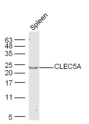 Sample: Spleen (Mouse) Lysate at 40 ug
Primary: Anti-CLEC5A(SL2663R) at 1/300 dilution
Secondary: IRDye800CW Goat Anti-Rabbit IgG at 1/20000 dilution
Predicted band size: 22 kD
Observed band size: 22 kD
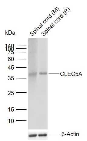 Sample:
Lane 1: Mouse Spinal cord tissue lysates
Lane 2: Rat Spinal cord tissue lysates
Primary: Anti-CLEC5A (SL2663R) at 1/1000 dilution
Secondary: IRDye800CW Goat Anti-Rabbit IgG at 1/20000 dilution
Predicted band size: 22 kDa
Observed band size: 37 kDa
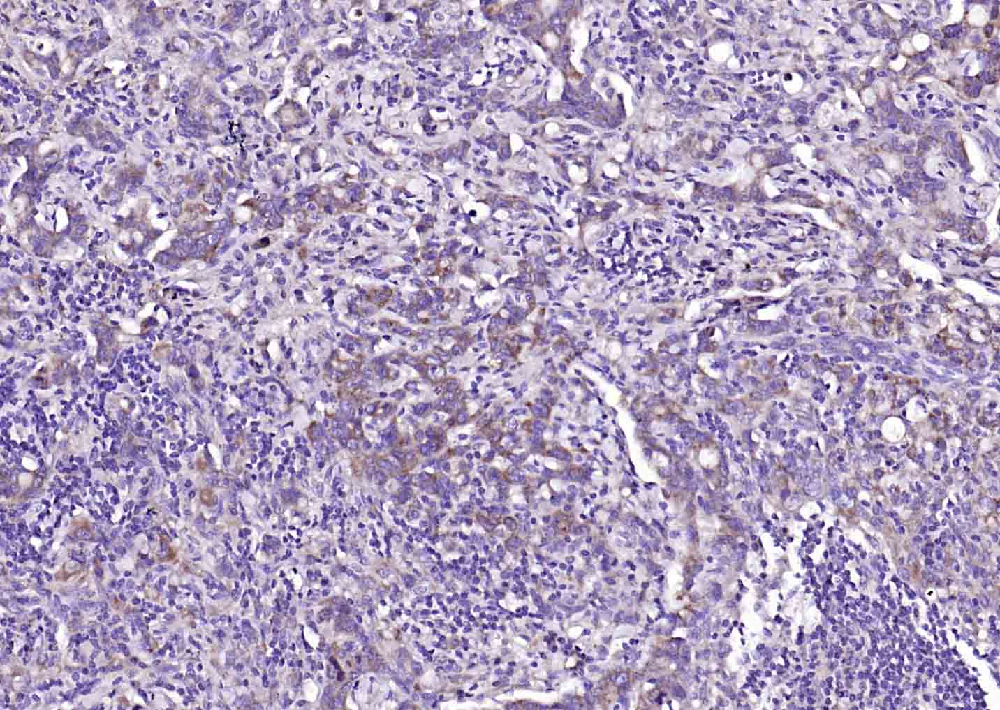 Paraformaldehyde-fixed, paraffin embedded (human gastric carcinoma); Antigen retrieval by boiling in sodium citrate buffer (pH6.0) for 15min; Block endogenous peroxidase by 3% hydrogen peroxide for 20 minutes; Blocking buffer (normal goat serum) at 37°C for 30min; Antibody incubation with (CLEC5A) Polyclonal Antibody, Unconjugated (SL2663R) at 1:200 overnight at 4°C, followed by operating according to SP Kit(Rabbit) (sp-0023) instructionsand DAB staining.
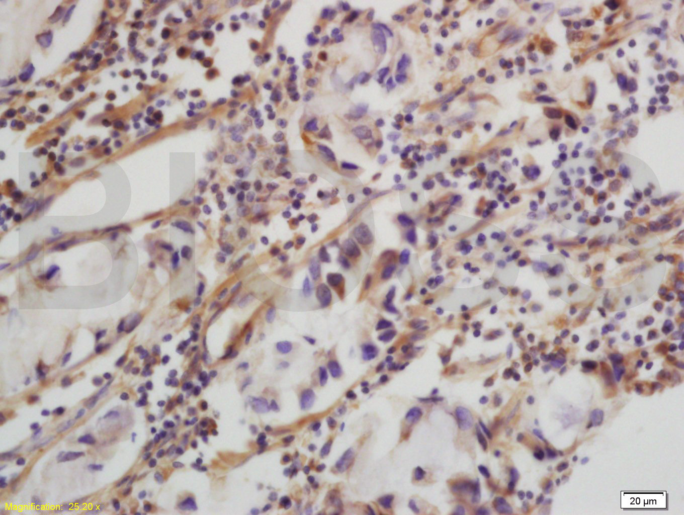 Tissue/cell: human colon carcinoma; 4% Paraformaldehyde-fixed and paraffin-embedded;
Antigen retrieval: citrate buffer ( 0.01M, pH 6.0 ), Boiling bathing for 15min; Block endogenous peroxidase by 3% Hydrogen peroxide for 30min; Blocking buffer (normal goat serum,C-0005) at 37℃ for 20 min;
Incubation: Anti-CLEC5A/MDL1 Polyclonal Antibody, Unconjugated(SL2663R) 1:200, overnight at 4°C, followed by conjugation to the secondary antibody(SP-0023) and DAB(C-0010) staining
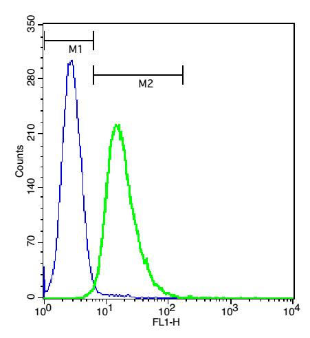 Cell: U937.
Concentration: 1:100.
Incubation: 40 minutes, room temperature.
Host/Blank: U937 cells.
Flow cytometric analysis of Rabbit Anti-CLEC5A antibody (SL2663R) (green) compared with control in the absence of primary antibody (blue) followed by U937 cells.
secondary antibody: Goat Anti-rabbit IgG/FITC antibody (SL0295G-FITC)
Cartpieces
Totalgoods,subtotals:¥Checkout
Partial purchase records(bought amounts latest0)
No one bought this product
User Comment(Total0User Comment Num)
- No comment


 +86 571 56623320


