
Rabbit Anti-LAMP2 antibody
CD107 antigen-like family member B; CD107b; CD107b antigen; Igp110; Igp2; LAMP 2; LAMP 2C; LAMP B; LAMP-2; LAMP2_RAT; LAMPB; LGP110; Lysosomal associated membrane protein 2C; Lysosome associated membrane glycoprotein 2 precursor; Lysosome associated membr
View History [Clear]
Details
Product Name LAMP2 Chinese Name 溶酶体相关膜蛋白2(CD107B)抗体 Alias CD107 antigen-like family member B; CD107b; CD107b antigen; Igp110; Igp2; LAMP 2; LAMP 2C; LAMP B; LAMP-2; LAMP2_RAT; LAMPB; LGP110; Lysosomal associated membrane protein 2C; Lysosome associated membrane glycoprotein 2 precursor; Lysosome associated membrane protein 2; Lysosome-associated membrane glycoprotein 2; Lysosome-associated membrane protein 2; MAC3. literatures Research Area Tumour Cardiovascular Cell biology Signal transduction Stem cells The new supersedes the old Mitochondrion Immunogen Species Rabbit Clonality Polyclonal React Species Human, Mouse, Rat, (predicted: Dog, Pig, Cow, Horse, Rabbit, ) Applications WB=1:500-2000 ELISA=1:5000-10000 IHC-P=1:100-500 IHC-F=1:100-500 IF=1:100-500 (Paraffin sections need antigen repair)
not yet tested in other applications.
optimal dilutions/concentrations should be determined by the end user.Theoretical molecular weight 45kDa Detection molecular weight 110 kDa Cellular localization cytoplasmic The cell membrane Mitochondrion Form Liquid Concentration 1mg/ml immunogen KLH conjugated synthetic peptide derived from human LAMP2: 301-411/411 Lsotype IgG Purification affinity purified by Protein A Buffer Solution 0.01M TBS(pH7.4) with 1% BSA, 0.03% Proclin300 and 50% Glycerol. Storage Shipped at 4℃. Store at -20 °C for one year. Avoid repeated freeze/thaw cycles. Attention This product as supplied is intended for research use only, not for use in human, therapeutic or diagnostic applications. PubMed PubMed Product Detail The protein encoded by the LAMP2 gene is a member of a family of membrane glycoproteins. This glycoprotein provides selectins with carbohydrate ligands and is thought to play a role in tumor cell metastasis. It may also function in the protection, maintenance, and adhesion of the lysosome. Alternative splicing of the gene produces two known products, LAMP2a and LAMP2b. Isoform LAMP2a is highly expressed in placenta, lung and liver and has low expression in brain and skeletal muscle. The isoform detected by this antibody, LAMP2b, is highly expressed in skeletal muscle less so in brain, placental, lung, kidney and pancreas and has very low expression in liver.
Function:
Implicated in tumor cell metastasis. May function in protection of the lysosomal membrane from autodigestion, maintenance of the acidic environment of the lysosome, adhesion when expressed on the cell surface (plasma membrane), and inter-and intracellular signal transduction. Protects cells from the toxic effects of methylating mutagens.
Subcellular Location:
Cell membrane; Single-pass type I membrane protein. Endosome membrane; Single-pass type I membrane protein. Lysosome membrane; Single-pass type I membrane protein. Note=This protein shuttles between lysosomes, endosomes, and the plasma membrane.
Post-translational modifications:
O- and N-glycosylated; some of the N-glycans attached to Lamp-2 are polylactosaminoglycans.
Similarity:
Belongs to the LAMP family.
SWISS:
P13473
Gene ID:
3920
Database links:
Entrez Gene: 3920 Human
Entrez Gene: 16784 Mouse
Omim: 309060 Human
SwissProt: P13473 Human
SwissProt: P17047 Mouse
Unigene: 496684 Human
Unigene: 486 Mouse
Product Picture 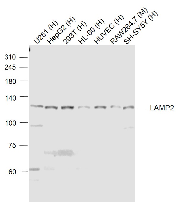 Sample:
Lane 1: U251 (Human) Cell Lysate at 30 ug
Lane 2: HepG2 (Human) Cell Lysate at 30 ug
Lane 3: 293T (Human) Cell Lysate at 30 ug
Lane 4: HL-60 (Human) Cell Lysate at 30 ug
Lane 5: HUVEC (Human) Cell Lysate at 30 ug
Lane 6: RAW264.7 (Mouse) Cell Lysate at 30 ug
Lane 7: SH-SY5Y (Human) Cell Lysate at 30 ug
Primary: Anti-LAMP2 (SL2379R) at 1/1000 dilution
Secondary: IRDye800CW Goat Anti-Rabbit IgG at 1/20000 dilution
Predicted band size: 120 kD
Observed band size: 120 kD
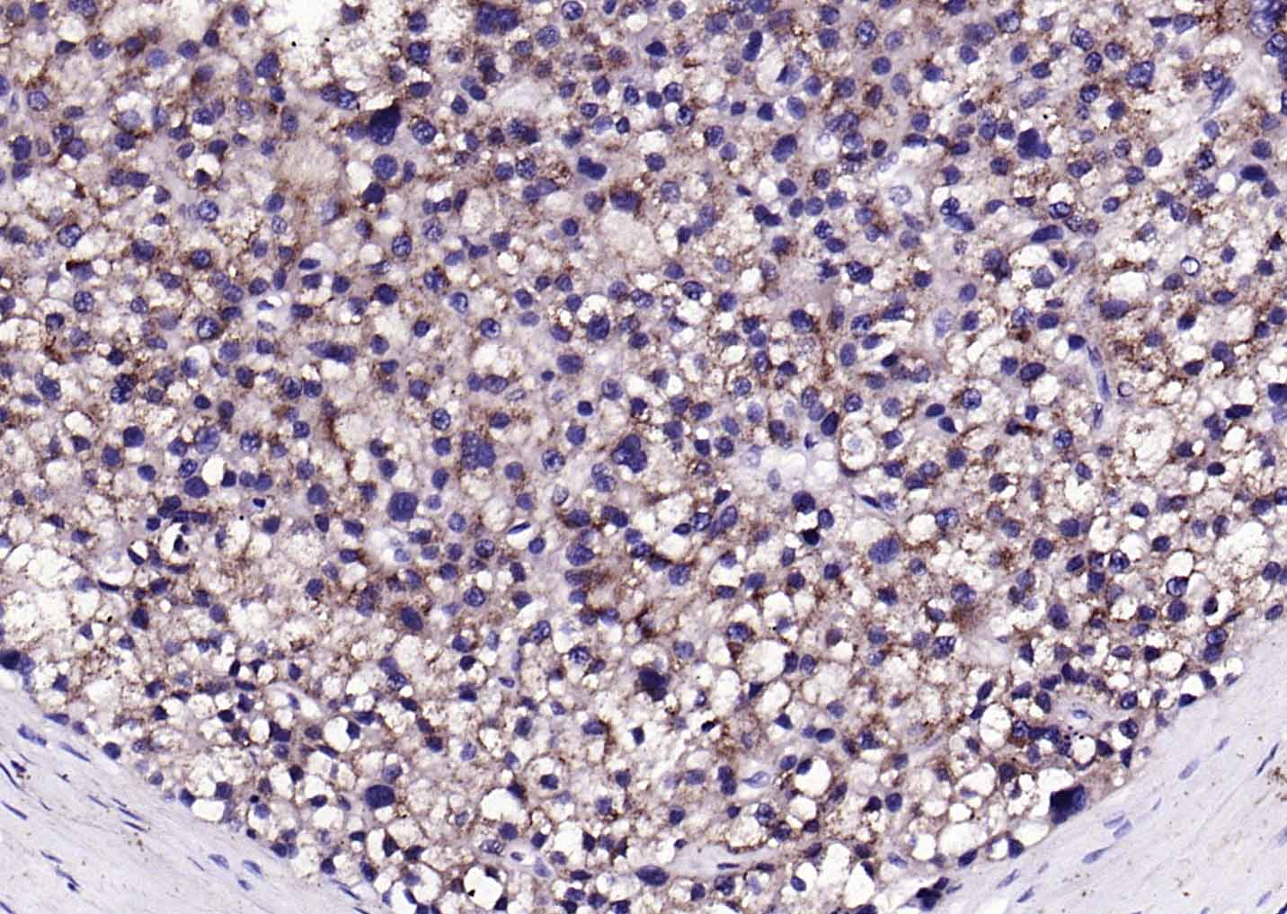 Paraformaldehyde-fixed, paraffin embedded (human liver carcinoma); Antigen retrieval by boiling in sodium citrate buffer (pH6.0) for 15min; Block endogenous peroxidase by 3% hydrogen peroxide for 20 minutes; Blocking buffer (normal goat serum) at 37°C for 30min; Antibody incubation with (LAMP2) Polyclonal Antibody, Unconjugated (SL2379R) at 1:200 overnight at 4°C, followed by operating according to SP Kit(Rabbit) (sp-0023) instructionsand DAB staining.
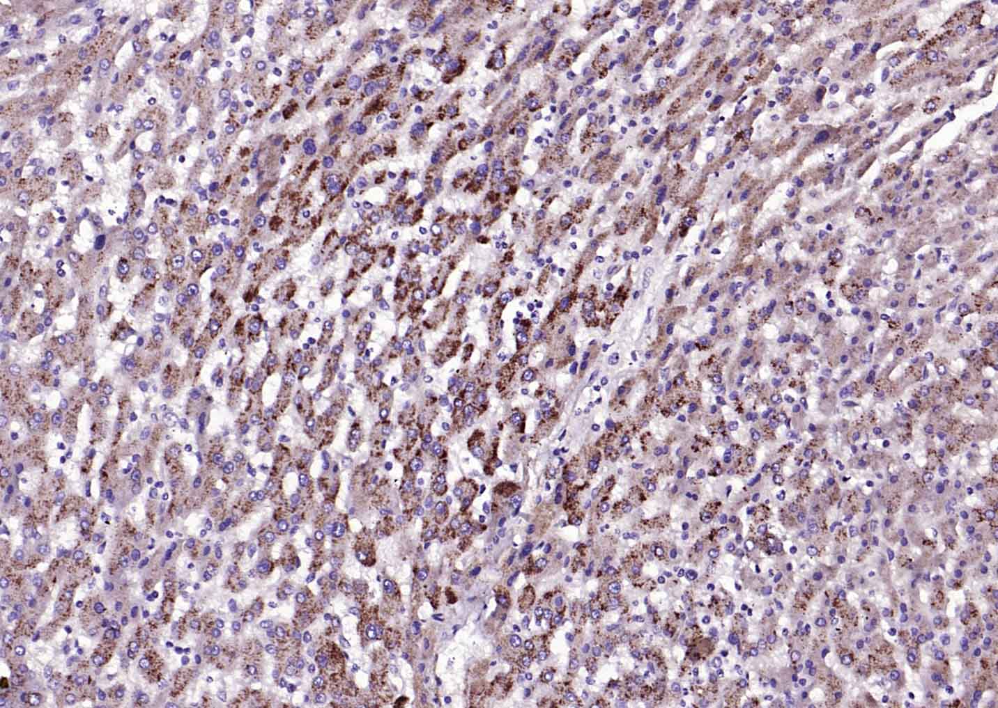 Paraformaldehyde-fixed, paraffin embedded (human liver ); Antigen retrieval by boiling in sodium citrate buffer (pH6.0) for 15min; Block endogenous peroxidase by 3% hydrogen peroxide for 20 minutes; Blocking buffer (normal goat serum) at 37°C for 30min; Antibody incubation with (LAMP2) Polyclonal Antibody, Unconjugated (SL2379R) at 1:200 overnight at 4°C, followed by operating according to SP Kit(Rabbit) (sp-0023) instructionsand DAB staining.
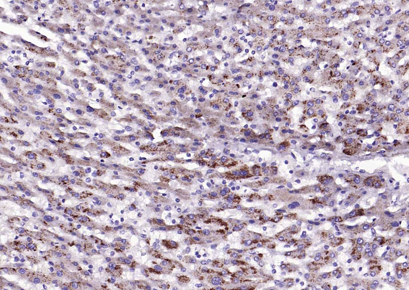 Paraformaldehyde-fixed, paraffin embedded (human liver ); Antigen retrieval by boiling in sodium citrate buffer (pH6.0) for 15min; Block endogenous peroxidase by 3% hydrogen peroxide for 20 minutes; Blocking buffer (normal goat serum) at 37°C for 30min; Antibody incubation with (LAMP2) Polyclonal Antibody, Unconjugated (SL2379R) at 1:200 overnight at 4°C, followed by operating according to SP Kit(Rabbit) (sp-0023) instructionsand DAB staining.
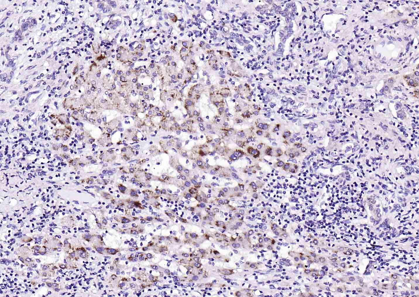 Paraformaldehyde-fixed, paraffin embedded (human liver carcinoma); Antigen retrieval by boiling in sodium citrate buffer (pH6.0) for 15min; Block endogenous peroxidase by 3% hydrogen peroxide for 20 minutes; Blocking buffer (normal goat serum) at 37°C for 30min; Antibody incubation with (LAMP2) Polyclonal Antibody, Unconjugated (SL2379R) at 1:200 overnight at 4°C, followed by operating according to SP Kit(Rabbit) (sp-0023) instructionsand DAB staining.
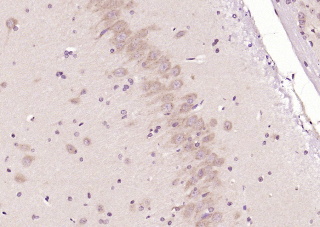 Paraformaldehyde-fixed, paraffin embedded (rat brain); Antigen retrieval by boiling in sodium citrate buffer (pH6.0) for 15min; Block endogenous peroxidase by 3% hydrogen peroxide for 20 minutes; Blocking buffer (normal goat serum) at 37°C for 30min; Antibody incubation with (LAMP2) Polyclonal Antibody, Unconjugated (SL2379R) at 1:200 overnight at 4°C, followed by operating according to SP Kit(Rabbit) (sp-0023) instructionsand DAB staining.
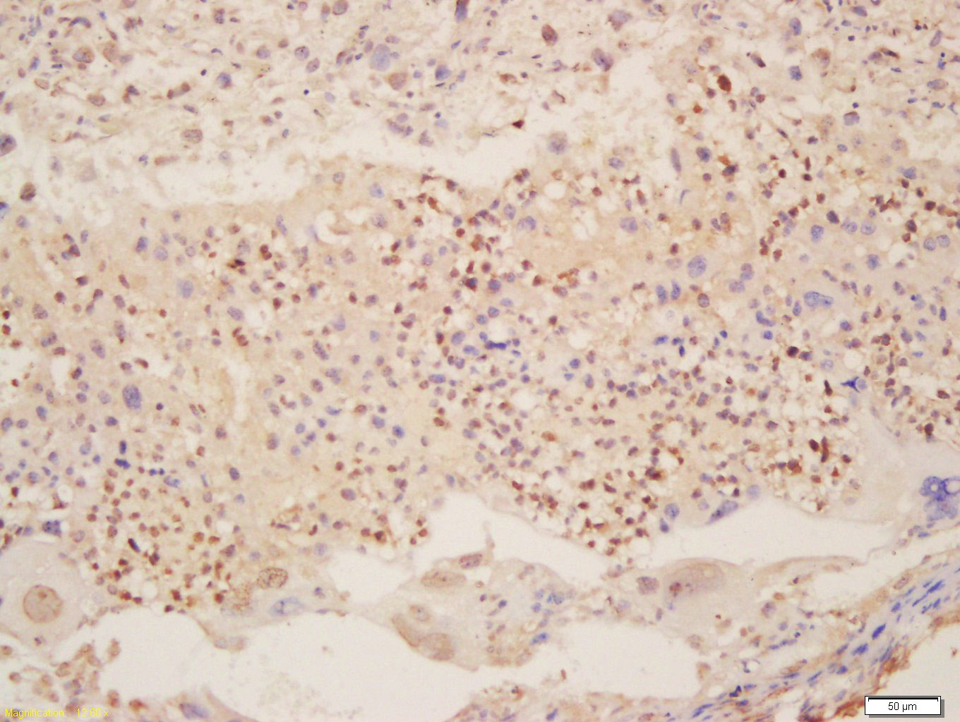 Tissue/cell: mouse placenta tissue; 4% Paraformaldehyde-fixed and paraffin-embedded;
Antigen retrieval: citrate buffer ( 0.01M, pH 6.0 ), Boiling bathing for 15min; Block endogenous peroxidase by 3% Hydrogen peroxide for 30min; Blocking buffer (normal goat serum,C-0005) at 37℃ for 20 min;
Incubation: Anti-LAMP2 Polyclonal Antibody, Unconjugated(SL2397R) 1:200, overnight at 4°C, followed by conjugation to the secondary antibody(SP-0023) and DAB(C-0010) staining
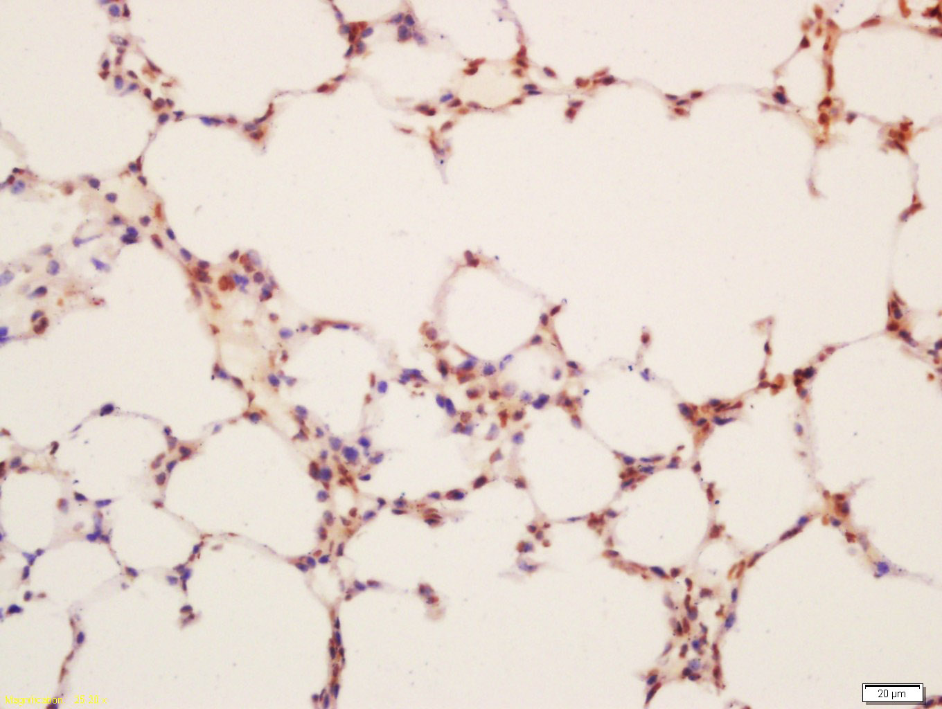 Tissue/cell: mouse lung tissue; 4% Paraformaldehyde-fixed and paraffin-embedded;
Antigen retrieval: citrate buffer ( 0.01M, pH 6.0 ), Boiling bathing for 15min; Block endogenous peroxidase by 3% Hydrogen peroxide for 30min; Blocking buffer (normal goat serum,C-0005) at 37℃ for 20 min;
Incubation: Anti-LAMP2 Polyclonal Antibody, Unconjugated(SL2397R) 1:200, overnight at 4°C, followed by conjugation to the secondary antibody(SP-0023) and DAB(C-0010) staining
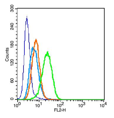 Blank control(blue):U937 (fixed with 2% paraformaldehyde (10 min)).
Primary Antibody:Rabbit Anti- LAMP2 antibody(SL2379R), Dilution: 1μg in 100 μL 1X PBS containing 0.5% BSA;
Isotype Control Antibody: Rabbit IgG(orange) ,used under the same conditions );
Secondary Antibody: Goat anti-rabbit IgG-PE(white blue), Dilution: 1:200 in 1 X PBS containing 0.5% BSA.
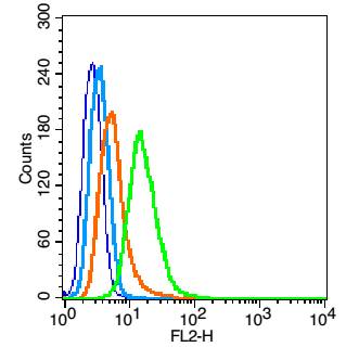 Blank control(blue): HELA (fixed with 2% paraformaldehyde (10 min), then permeabilized with 90% ice-cold methanol for 30 min on ice).
Primary Antibody:Rabbit Anti-LAMP2 Receptor beta antibody(SL2379R), Dilution: 5μg in 100 μL 1X PBS containing 0.5% BSA;
Isotype Control Antibody: Rabbit IgG(orange) ,used under the same conditions );
Secondary Antibody: Goat anti-rabbit IgG-PE(white blue), Dilution: 1:200 in 1 X PBS containing 0.5% BSA.
Cartpieces
Totalgoods,subtotals:¥Checkout
Bought notes(bought amounts latest0)
No one bought this product
User Comment(Total0User Comment Num)
- No comment


 +86 571 56623320
 +86 18668110335

