
Rabbit Anti-ITGAV antibody
Integrin alpha-V light chain; Integrin Alpha V; CD 51; CD51; CD51 antigen; HGNC; Msk 8; Msk8; Vitronectin receptor alpha polypeptide; Vitronectin receptor alpha polypeptide antigen CD51; Vitronectin receptor subunit alpha; VNRA; VTNR; ITAV_HUMAN; Integrin
View History [Clear]
Details
Product Name ITGAV Chinese Name 整合素αV(CD51)抗体 Alias Integrin alpha-V light chain; Integrin Alpha V; CD 51; CD51; CD51 antigen; HGNC; Msk 8; Msk8; Vitronectin receptor alpha polypeptide; Vitronectin receptor alpha polypeptide antigen CD51; Vitronectin receptor subunit alpha; VNRA; VTNR; ITAV_HUMAN; Integrin alpha-V Precursor. literatures Research Area Cardiovascular Signal transduction The cell membrane受体 Cell adhesion molecule Immunogen Species Rabbit Clonality Polyclonal React Species Human, Mouse, (predicted: Rat, Chicken, Dog, Pig, Cow, Horse, ) Applications WB=1:1000-10000 ELISA=1:5000-10000 IHC-P=1:100-500 IHC-F=1:100-500 Flow-Cyt=1μg/Test ICC=1:100 IF=1:100-500 (Paraffin sections need antigen repair)
not yet tested in other applications.
optimal dilutions/concentrations should be determined by the end user.Theoretical molecular weight 95/113kDa Cellular localization The cell membrane Form Liquid Concentration 1mg/ml immunogen KLH conjugated synthetic peptide derived from human Integrin alpha V: 51-150/1048 <Extracellular> Lsotype IgG Purification affinity purified by Protein A Buffer Solution 0.01M TBS(pH7.4) with 1% BSA, 0.03% Proclin300 and 50% Glycerol. Storage Shipped at 4℃. Store at -20 °C for one year. Avoid repeated freeze/thaw cycles. Attention This product as supplied is intended for research use only, not for use in human, therapeutic or diagnostic applications. PubMed PubMed Product Detail The product of this gene belongs to the integrin alpha chain family. Integrins are heterodimeric integral membrane proteins composed of an alpha subunit and a beta subunit that function in cell surface adhesion and signaling. The encoded preproprotein is proteolytically processed to generate light and heavy chains that comprise the alpha V subunit. This subunit associates with beta 1, beta 3, beta 5, beta 6 and beta 8 subunits. The heterodimer consisting of alpha V and beta 3 subunits is also known as the vitronectin receptor. This integrin may regulate angiogenesis and cancer progression. Alternative splicing results in multiple transcript variants. Note that the integrin alpha 5 and integrin alpha V subunits are encoded by distinct genes. [provided by RefSeq, Oct 2015]
Function:
The alpha-V integrins are receptors for vitronectin, cytotactin, fibronectin, fibrinogen, laminin, matrix metalloproteinase-2, osteopontin, osteomodulin, prothrombin, thrombospondin and vWF. They recognize the sequence R-G-D in a wide array of ligands. In case of HIV-1 infection, the interaction with extracellular viral Tat protein seems to enhance angiogenesis in Kaposi's sarcoma lesions.
Subunit:
Heterodimer of an alpha and a beta subunit. The alpha subunit is composed of an heavy and a light chain linked by a disulfide bond. Alpha-V associates with either beta-1, beta-3, beta-5, beta-6 or beta-8 subunit. Interacts with HIV-1 Tat. Alpha-V/beta-6 binds to foot-and-mouth disease virus (FMDV) VP1 protein and acts as a receptor for this viru. Alpha-V/beta-6 binds to coxsackievirus A9 and coxsackievirus B1 capsid proteins and acts as a receptor for these viruses. Interacts with RAB25.
Subcellular Location:
Membrane; Single-pass type I membrane protein.
Similarity:
Belongs to the integrin alpha chain family.
Contains 7 FG-GAP repeats.
SWISS:
P06756
Gene ID:
3685
Database links:Entrez Gene: 3685 Human
Entrez Gene: 16410 Mouse
SwissProt: P06756 Human
SwissProt: P43406 Mouse
Product Picture 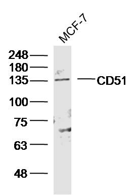 Sample:MCF-7 Cell (Human) Lysate at 40 ug
Primary: Anti- CD51 (SL2250R) at 1/300 dilution
Secondary: IRDye800CW Goat Anti-Rabbit IgG at 1/20000 dilution
Predicted band size: 95/113 kD
Observed band size: 135 kD
 Sample:
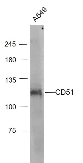
A549(Human) Cell Lysate at 30 ug
Primary: Anti- CD51 (SL2250R) at 1/1000 dilution
Secondary: IRDye800CW Goat Anti-Rabbit IgG at 1/20000 dilution
Predicted band size: 95/113 kD
Observed band size: 113 kD
 Sample:
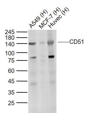
Lane 1: A549 (Human) Cell Lysate at 30 ug
Lane 2: MCF-7 (Human) Cell Lysate at 30 ug
Lane 3: Huvec (Human) Cell Lysate at 30 ug
Primary: Anti-CD51 (SL2250R) at 1/1000 dilution
Secondary: IRDye800CW Goat Anti-Rabbit IgG at 1/20000 dilution
Predicted band size: 125-140 kD
Observed band size: 140 kD
 Sample:
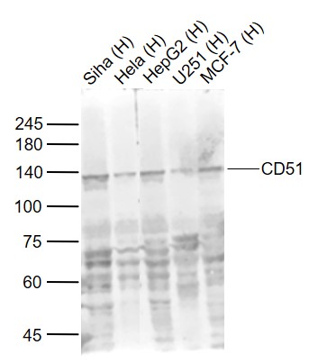
Lane 1: Siha (Human) Cell Lysate at 30 ug
Lane 2: Hela (Human) Cell Lysate at 30 ug
Lane 3: HepG2 (Human) Cell Lysate at 30 ug
Lane 4: U251 (Human) Cell Lysate at 30 ug
Lane 5: MCF-7 (Human) Cell Lysate at 30 ug
Primary: Anti-CD51 (SL2250R) at 1/1000 dilution
Secondary: IRDye800CW Goat Anti-Rabbit IgG at 1/20000 dilution
Predicted band size: 125-140 kD
Observed band size: 135 kD
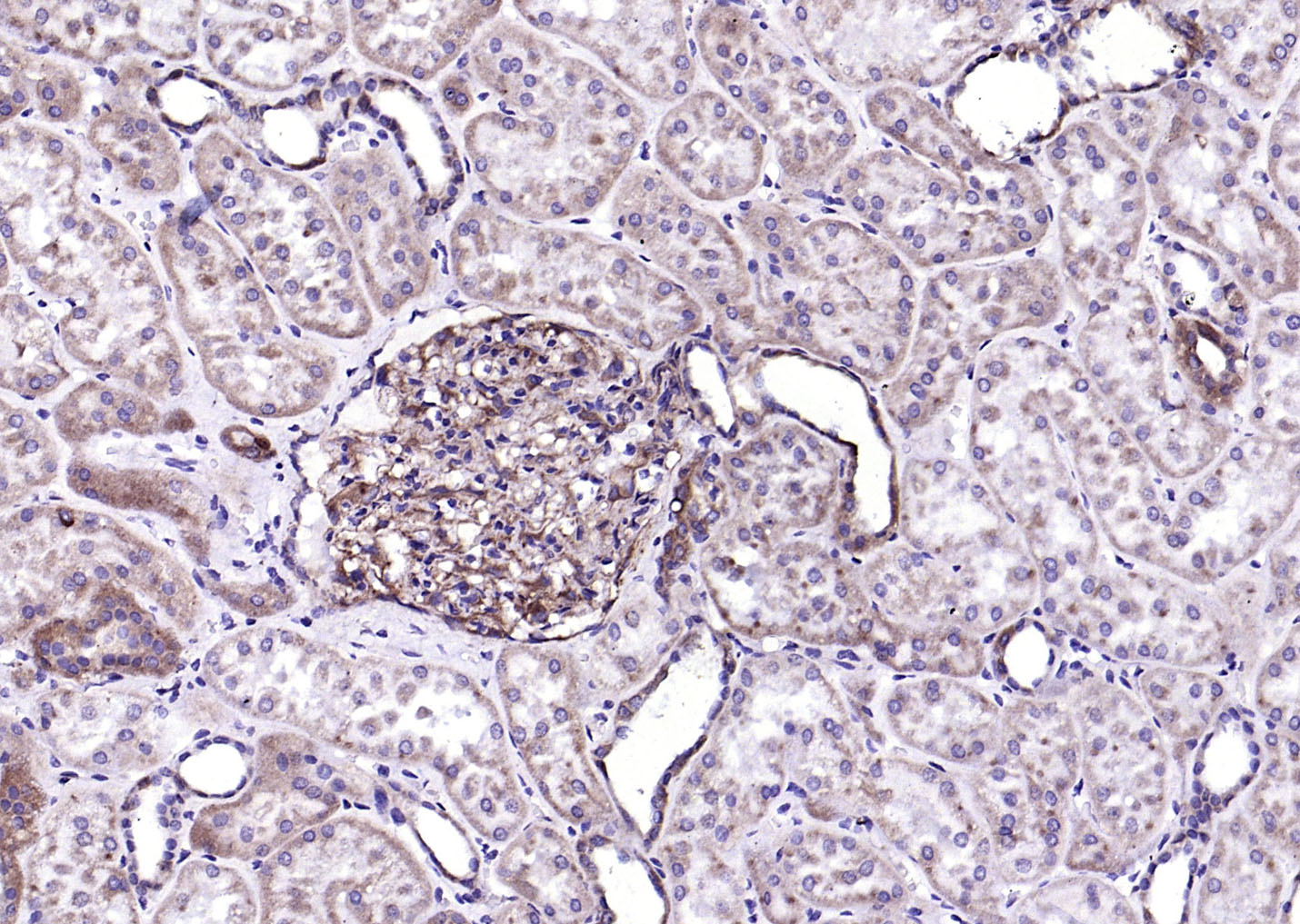 Paraformaldehyde-fixed, paraffin embedded (human kidney); Antigen retrieval by boiling in sodium citrate buffer (pH6.0) for 15min; Block endogenous peroxidase by 3% hydrogen peroxide for 20 minutes; Blocking buffer (normal goat serum) at 37°C for 30min; Antibody incubation with (ITGAV) Polyclonal Antibody, Unconjugated (SL2250R) at 1:500 overnight at 4°C, followed by operating according to SP Kit(Rabbit) (sp-0023) instructionsand DAB staining.
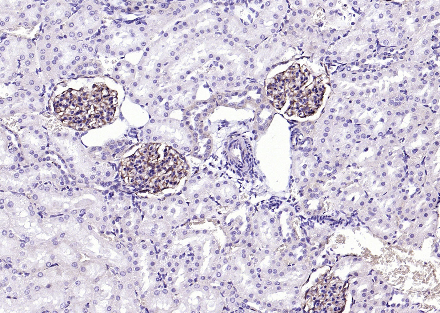 Paraformaldehyde-fixed, paraffin embedded (rat kidney); Antigen retrieval by boiling in sodium citrate buffer (pH6.0) for 15min; Block endogenous peroxidase by 3% hydrogen peroxide for 20 minutes; Blocking buffer (normal goat serum) at 37°C for 30min; Antibody incubation with (ITGAV) Polyclonal Antibody, Unconjugated (SL2250R) at 1:500 overnight at 4°C, followed by operating according to SP Kit(Rabbit) (sp-0023) instructionsand DAB staining.
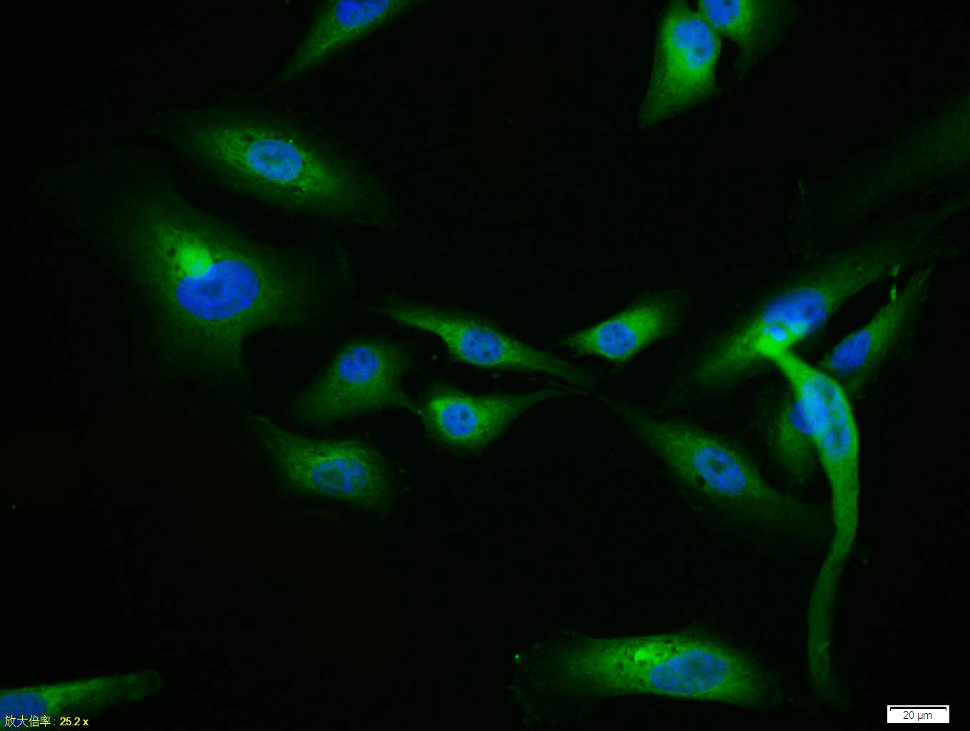 U-2OS cell; 4% Paraformaldehyde-fixed; Triton X-100 at room temperature for 20 min; Blocking buffer (normal goat serum, C-0005) at 37°C for 20 min; Antibody incubation with (CD51) polyclonal Antibody, Unconjugated (SL2250R) 1:100, 90 minutes at 37°C; followed by a conjugated Goat Anti-Rabbit IgG antibody at 37°C for 90 minutes, DAPI (blue, C02-04002) was used to stain the cell nuclei.
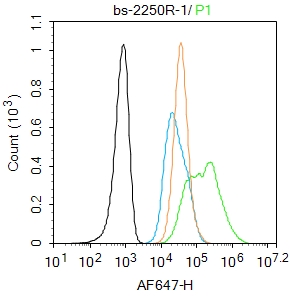 Blank control:U2OS.
Primary Antibody (green line): Rabbit Anti-CD51 antibody (SL2250R)
Dilution: 1μg /10^6 cells;
Isotype Control Antibody (orange line): Rabbit IgG .
Secondary Antibody : Goat anti-rabbit IgG-AF647
Dilution: 1μg /test.
Protocol
The cells were fixed with 4% PFA (10min at room temperature)and then permeabilized with 0.1%PBST for 20 min at room temperature. The cells were then incubated in 5%BSA to block non-specific protein-protein interactions for 30 min at room temperature .Cells stained with Primary Antibody for 30 min at room temperature. The secondary antibody used for 40 min at room temperature. Acquisition of 20,000 events was performed.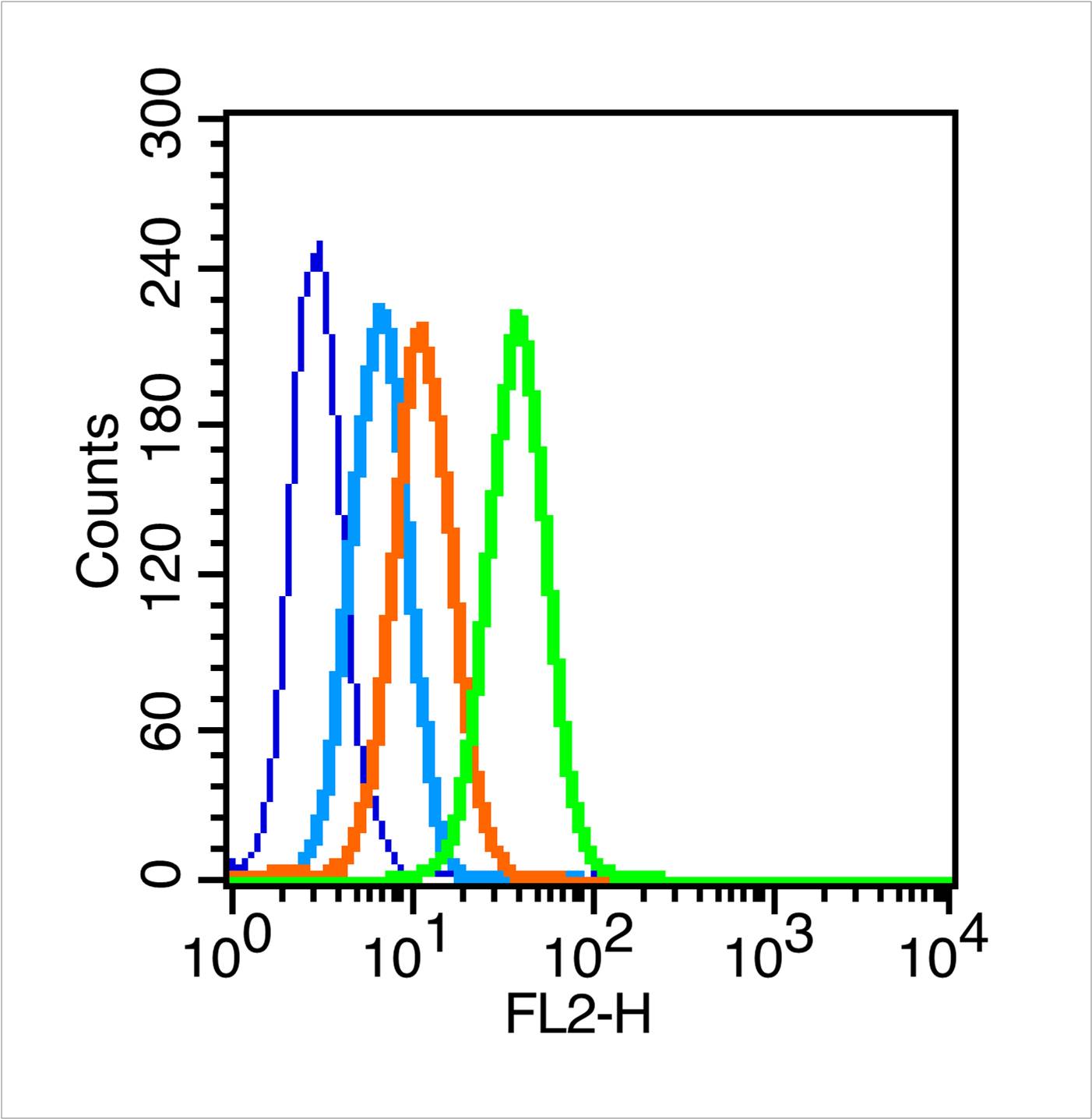 Blank control (blue line): MCF7 (fixed with 70% methanol overnight at 4℃).
Primary Antibody (green line): Rabbit Anti-CD51 antibody (SL2250R), Dilution: 1μg /10^6 cells;
Isotype Control Antibody (orange line): Rabbit IgG .
Secondary Antibody (white blue line): Goat anti-rabbit IgG-PE,Dilution: 1μg /test.
Cartpieces
Totalgoods,subtotals:¥Checkout
References (0)
No References
Bought notes(bought amounts latest0)
No one bought this product
User Comment(Total0User Comment Num)
- No comment


 +86 571 56623320
 +86 18668110335

