
Rabbit Anti-N-cadherin antibody
Cadherin 2; Cadherin 2 N cadherin neuronal; Cadherin 2 type 1; Cadherin 2 type 1 N cadherin neuronal; cadherin 2 type 1 N-cadherin neuronal; Cadherin2; Calcium dependent adhesion protein neuronal; CD325; CD325 antigen; CDH2; CDHN; CDw325; CDw325 antigen;
View History [Clear]
Details
Product Name N-cadherin Chinese Name N-钙粘附分子抗体 Alias Cadherin 2; Cadherin 2 N cadherin neuronal; Cadherin 2 type 1; Cadherin 2 type 1 N cadherin neuronal; cadherin 2 type 1 N-cadherin neuronal; Cadherin2; Calcium dependent adhesion protein neuronal; CD325; CD325 antigen; CDH2; CDHN; CDw325; CDw325 antigen; N cadherin 1; NCAD; Neural Cadherin; Neural Cadherin. literatures Research Area Tumour Cell biology immunology Kinases and Phosphatases Cell adhesion molecule Immunogen Species Rabbit Clonality Polyclonal React Species Human, Mouse, Cow, (predicted: Rat, Pig, ) Applications WB=1:500-2000 ELISA=1:5000-10000 IHC-P=1:100-500 IHC-F=1:100-500 Flow-Cyt=1:100 ICC=1:100 IF=1:100-500 (Paraffin sections need antigen repair)
not yet tested in other applications.
optimal dilutions/concentrations should be determined by the end user.Theoretical molecular weight 100kDa Cellular localization The cell membrane Form Liquid Concentration 1mg/ml immunogen KLH conjugated synthetic peptide derived from human N-cadherin: 701-800/905 Lsotype IgG Purification affinity purified by Protein A Buffer Solution 0.01M TBS(pH7.4) with 1% BSA, 0.03% Proclin300 and 50% Glycerol. Storage Shipped at 4℃. Store at -20 °C for one year. Avoid repeated freeze/thaw cycles. Attention This product as supplied is intended for research use only, not for use in human, therapeutic or diagnostic applications. PubMed PubMed Product Detail This gene is a classical cadherin from the cadherin superfamily. The encoded protein is a calcium dependent cell-cell adhesion glycoprotein comprised of five extracellular cadherin repeats, a transmembrane region and a highly conserved cytoplasmic tail. The protein functions during gastrulation and is required for establishment of left-right asymmetry. At certain central nervous system synapses, presynaptic to postsynaptic adhesion is mediated at least in part by this gene product.
Function:
Cadherins are calcium dependent cell adhesion proteins. They preferentially interact with themselves in a homophilic manner in connecting cells; cadherins may thus contribute to the sorting of heterogeneous cell types. CDH2 may be involved in neuronal recognition mechanism. In hippocampal neurons, may regulate dendritic spine density.
Subcellular Location:
Cell membrane.
Similarity:
Contains 5 cadherin domains.
SWISS:
P19022
Gene ID:
1000
Database links:Entrez Gene: 1000 Human
Entrez Gene: 12558 Mouse
Omim: 114020 Human
SwissProt: P19022 Human
SwissProt: P15116 Mouse
Unigene: 464829 Human
Unigene: 606106 Human
Unigene: 257437 Mouse
Unigene: 23200 Rat
细胞粘附蛋白(Call Adhesion Protein)
N-钙粘附分子属于依赖钙离子的细胞间glycoprotein粘附分子的一部分,主要是参与细胞间同源性粘附,调控和介导细胞间相互反应。Product Picture 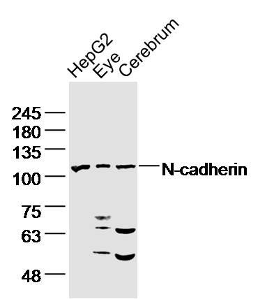 Sample:
HepG2 Cell Lysate at 40 ug
Eye (Mouse) Lysate at 40 ug
Cerebrum (Mouse) Lysate at 40 ug
Primary: Anti- N-cadherin (SL1172R)at 1/300 dilution
Secondary: IRDye800CW Goat Anti-Rabbit IgG at 1/20000 dilution
Predicted band size: 100kD
Observed band size: 110kD
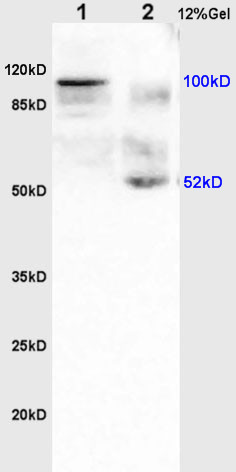 Sample:
Colon carcinoma(Human) lysate at 30ug;
Brain(Rat) lysate at 30ug;
Primary: Anti-CDH2/N-cadherin (SL1172R) at 1:200 dilution;
Secondary: HRP conjugated Goat Anti-Rabbit IgG(SL0295G-HRP) at 1: 3000 dilution;
Predicted band size : 100kD
Observed band size : 100kD
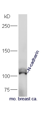 Sample:
Breast ca (Mouse) Lysate at 40 ug
Primary: Anti-N-cadherin (SL1172R) at 1/300 dilution
Secondary: IRDye800CW Goat Anti-Rabbit IgG at 1/20000 dilution
Predicted band size: 100 kD
Observed band size: 105 kD
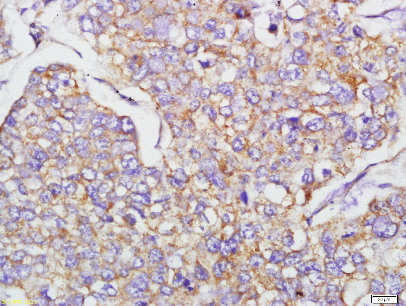 Tissue/cell: human lung carcinoma; 4% Paraformaldehyde-fixed and paraffin-embedded;
Antigen retrieval: citrate buffer ( 0.01M, pH 6.0 ), Boiling bathing for 15min; Block endogenous peroxidase by 3% Hydrogen peroxide for 30min; Blocking buffer (normal goat serum,C-0005) at 37℃ for 20 min;
Incubation: Anti-N-cadherin Polyclonal Antibody, Unconjugated(SL1172R) 1:400, overnight at 4°C, followed by conjugation to the secondary antibody(SP-0023) and DAB(C-0010) staining
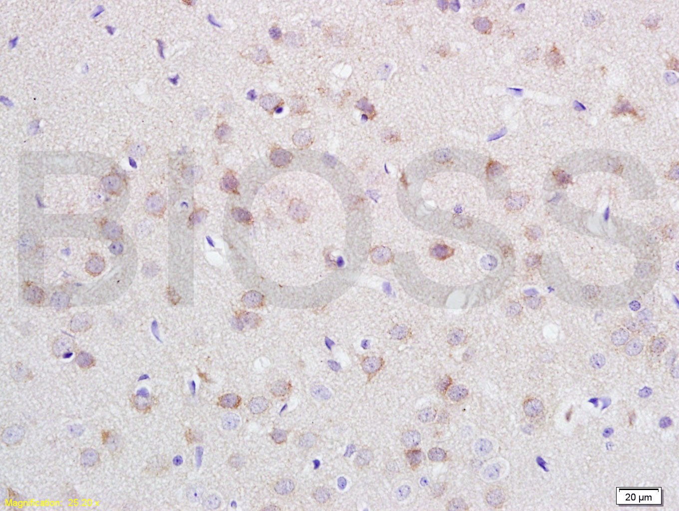 Tissue/cell: rat brain tissue; 4% Paraformaldehyde-fixed and paraffin-embedded;
Antigen retrieval: citrate buffer ( 0.01M, pH 6.0 ), Boiling bathing for 15min; Block endogenous peroxidase by 3% Hydrogen peroxide for 30min; Blocking buffer (normal goat serum,C-0005) at 37℃ for 20 min;
Incubation: Anti-N-cadherin Polyclonal Antibody, Unconjugated(SL1172R) 1:200, overnight at 4°C, followed by conjugation to the secondary antibody(SP-0023) and DAB(C-0010) staining
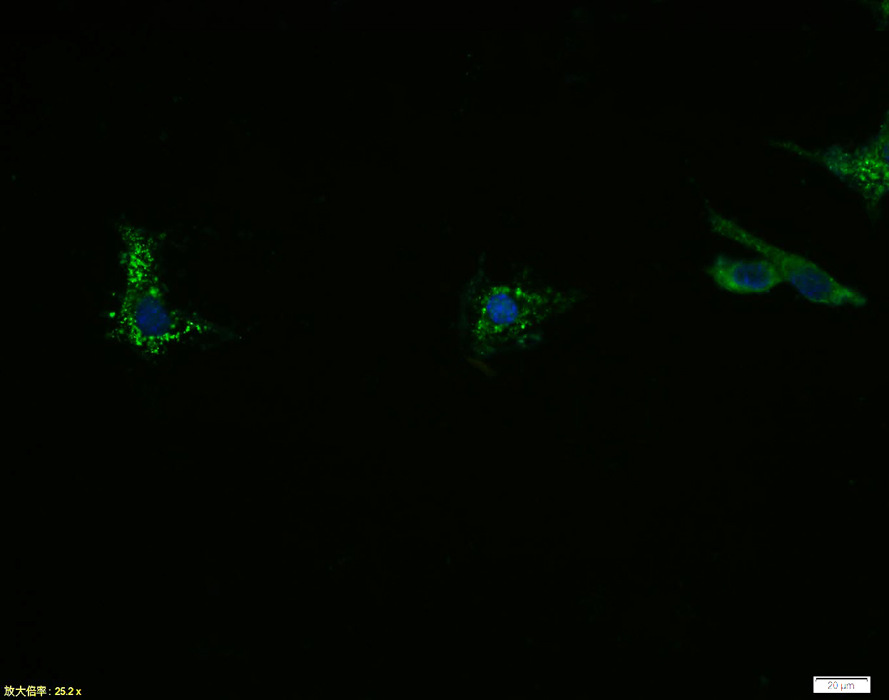 Tissue/cell:SH-SY5Y cell; 4% Paraformaldehyde-fixed; Triton X-100 at room temperature for 20 min; Blocking buffer (normal goat serum, C-0005) at 37°C for 20 min; Antibody incubation with (N-cadherin) polyclonal Antibody, Unconjugated (SL1172R) 1:100, 90 minutes at 37°C; followed by a FITC conjugated Goat Anti-Rabbit IgG antibody at 37°C for 90 minutes, DAPI (blue, C02-04002) was used to stain the cell nuclei.
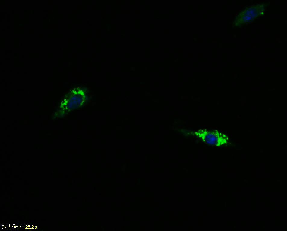 Tissue/cell:SH-SY5Y cell; 4% Paraformaldehyde-fixed; Triton X-100 at room temperature for 20 min; Blocking buffer (normal goat serum, C-0005) at 37°C for 20 min; Antibody incubation with (N-cadherin) polyclonal Antibody, Unconjugated (SL1172R) 1:100, 90 minutes at 37°C; followed by a FITC conjugated Goat Anti-Rabbit IgG antibody at 37°C for 90 minutes, DAPI (blue, C02-04002) was used to stain the cell nuclei.
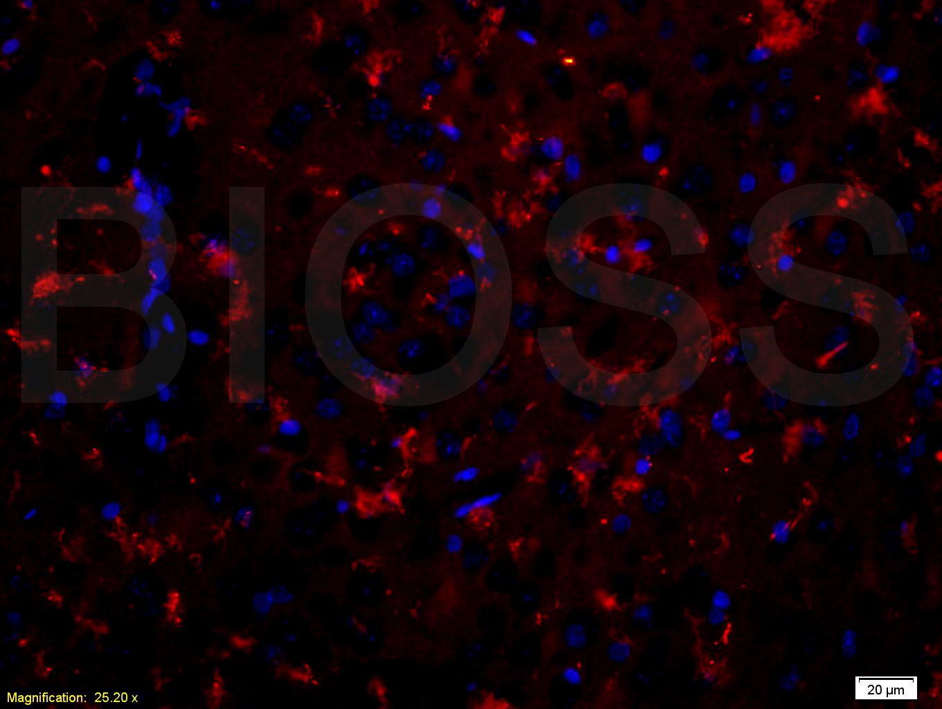 Tissue/cell: rat brain tissue;4% Paraformaldehyde-fixed and paraffin-embedded;
Antigen retrieval: citrate buffer ( 0.01M, pH 6.0 ), Boiling bathing for 15min; Blocking buffer (normal goat serum,C-0005) at 37℃ for 20 min;
Incubation: Anti-N-cadherin Polyclonal Antibody, Unconjugated(SL1172R) 1:200, overnight at 4°C; The secondary antibody was Goat Anti-Rabbit IgG, PE conjugated(SL0295G-PE)used at 1:200 dilution for 40 minutes at 37°C. DAPI(5ug/ml,blue,C-0033) was used to stain the cell nuclei
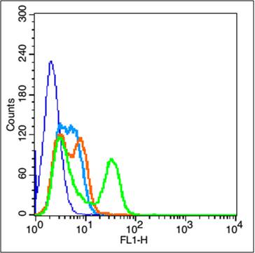 Blank control: Hepg2(blue).
Primary Antibody:Rabbit Anti-E cadherin antibody (SL1172R,Green); Dilution: 3μg in 100 μL 1X PBS containing 0.5% BSA;
Isotype Control Antibody: Rabbit IgG(orange) ,used under the same conditions;
Secondary Antibody: Goat anti-rabbit IgG-FITC(white blue), Dilution: 1:200 in 1 X PBS containing 0.5% BSA.
Protocol
The cells were fixed with 2% paraformaldehyde for 10 min at 37℃. Primary antibody (SL1172R, 3μg /1x10^6 cells) were incubated for 30 min at room temperature, followed by 1 X PBS containing 0.5% BSA + 1 0% goat serum (1 hour) to block non-specific protein-protein interactions. Then the Goat Anti-rabbit IgG/FITC antibody was added into the blocking buffer mentioned above to react with the primary antibody at 1/200 dilution for 40 min at room temperature. Acquisition of 20,000 events was performed.
Cartpieces
Totalgoods,subtotals:¥Checkout
Bought notes(bought amounts latest0)
No one bought this product
User Comment(Total0User Comment Num)
- No comment


 +86 571 56623320
 +86 18668110335

