
Rabbit Anti-HAS2 antibody
HA synthase 2; HAS2_HUMAN; Hyaluronan synthase 2; Hyaluronate synthase 2; Hyaluronic acid synthase 2.
View History [Clear]
Details
Product Name HAS2 Chinese Name 透明质酸合成酶2抗体 Alias HA synthase 2; HAS2_HUMAN; Hyaluronan synthase 2; Hyaluronate synthase 2; Hyaluronic acid synthase 2. literatures Research Area Cardiovascular Cell biology Developmental biology Neurobiology Signal transduction Cell adhesion molecule Immunogen Species Rabbit Clonality Polyclonal React Species Human, (predicted: Mouse, Rat, Chicken, Pig, Cow, Horse, Rabbit, Sheep, ) Applications WB=1:500-2000 ELISA=1:5000-10000 IHC-F=1:100-500 Flow-Cyt=1ug/Test IF=1:100-500 (Paraffin sections need antigen repair)
not yet tested in other applications.
optimal dilutions/concentrations should be determined by the end user.Theoretical molecular weight 64kDa Cellular localization The cell membrane Form Liquid Concentration 1mg/ml immunogen KLH conjugated synthetic peptide derived from human HAS2/Hyaluronan synthase 2: 401-500/552 <Extracellular> Lsotype IgG Purification affinity purified by Protein A Buffer Solution 0.01M TBS(pH7.4) with 1% BSA, 0.03% Proclin300 and 50% Glycerol. Storage Shipped at 4℃. Store at -20 °C for one year. Avoid repeated freeze/thaw cycles. Attention This product as supplied is intended for research use only, not for use in human, therapeutic or diagnostic applications. PubMed PubMed Product Detail Hyaluronan or hyaluronic acid (HA) is a high molecular weight unbranched polysaccharide synthesized by a wide variety of organisms from bacteria to mammals, and is a constituent of the extracellular matrix. It consists of alternating glucuronic acid and N-acetylglucosamine residues that are linked by beta-1-3 and beta-1-4 glycosidic bonds. HA is synthesized by membrane-bound synthase at the inner surface of the plasma membrane, and the chains are extruded through pore-like structures into the extracellular space. It serves a variety of functions, including space filling, lubrication of joints, and provision of a matrix through which cells can migrate. HA is actively produced during wound healing and tissue repair to provide a framework for ingrowth of blood vessels and fibroblasts. Changes in the serum concentration of HA are associated with inflammatory and degenerative arthropathies such as rheumatoid arthritis. In addition, the interaction of HA with the leukocyte receptor CD44 is important in tissue-specific homing by leukocytes, and overexpression of HA receptors has been correlated with tumor metastasis. HAS2 is a member of the newly identified vertebrate gene family encoding putative hyaluronan synthases, and its amino acid sequence shows significant homology to glycosaminoglycan synthetase (DG42) from Xenopus laevis, and human and murine hyaluronan synthase 1. [provided by RefSeq, Jul 2008]
Function:
Plays a role in hyaluronan/hyaluronic acid (HA) synthesis.
Subcellular Location:
Membrane.
Tissue Specificity:
Expressed in fibroblasts.
DISEASE:
Note=A chromosomal aberration involving HAS2 may be a cause of lipoblastomas, which are benign tumors resulting from transformation of adipocytes, usually diagnosed in children. 8q12.1 to 8q24.1 intrachromosomal rearrangement with PLAG1.
Similarity:
Belongs to the nodC/HAS family.
SWISS:
Q92819
Gene ID:
3037
Database links:Entrez Gene: 395594 Chicken
Entrez Gene: 100009708 Horse
Entrez Gene: 3037 Human
Entrez Gene: 15117 Mouse
Omim: 601636 Human
SwissProt: O57424 Chicken
SwissProt: Q92819 Human
SwissProt: P70312 Mouse
Unigene: 329 Chicken
Unigene: 159226 Human
Unigene: 5148 Mouse
Unigene: 87393 Rat
Product Picture 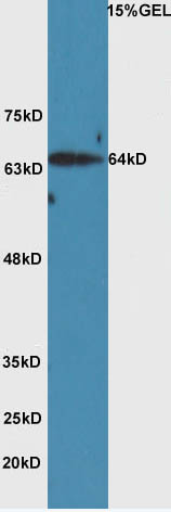 Sample: Huh7 Cell (Human) Lysate at 40 ug
Primary: Anti-HAS2 (SL11290R) at 1/300 dilution
Secondary: HRP conjugated Goat-Anti-rabbit IgG (SL0295G-HRP) at 1/5000 dilution
Predicted band size: 64 kD
Observed band size: 64 kD
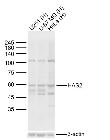 Sample:
Lane 1: Human U251 cell Lysates
Lane 2: Human U-87 MG cell Lysates
Lane 3: Human Hela cell Lysates
Primary: Anti-HAS2 (SL11290R) at 1/1000 dilution
Secondary: IRDye800CW Goat Anti-Rabbit IgG at 1/20000 dilution
Predicted band size: 64kDa
Observed band size: 60kDa
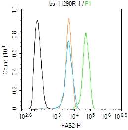 Blank control(black line):Hela.
Primary Antibody (green line): Rabbit Anti-HAS2 antibody (SL11290R)
Dilution:1ug/Test;
Secondary Antibody(white blue line): Goat anti-rabbit IgG-AF488
Dilution: 0.5ug/Test.
Isotype control(orange line): Normal Rabbit IgG
Protocol
The cells were fixed with 4% PFA (10min at room temperature)and then permeabilized with 90% ice-cold methanol for 20 min at -20℃, The cells were then incubated in 5%BSA to block non-specific protein-protein interactions for 30 min at room temperature .Cells stained with Primary Antibody for 30 min at room temperature. The secondary antibody used for 40 min at room temperature. Acquisition of 20,000 events was performed.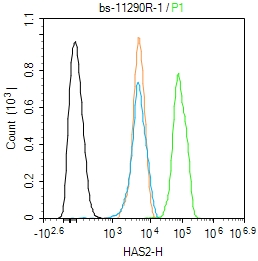 Blank control(black line):Hela.
Primary Antibody (green line): Rabbit Anti-HAS2 antibody (SL11290R)
Dilution:1ug/Test;
Secondary Antibody(white blue line): Goat anti-rabbit IgG-AF488
Dilution: 0.5ug/Test.
Isotype control(orange line): Normal Rabbit IgG
Protocol
The cells were fixed with 4% PFA (10min at room temperature)and then permeabilized with 90% ice-cold methanol for 20 min at -20℃, The cells were then incubated in 5%BSA to block non-specific protein-protein interactions for 30 min at room temperature .Cells stained with Primary Antibody for 30 min at room temperature. The secondary antibody used for 40 min at room temperature. Acquisition of 20,000 events was performed.
Cartpieces
Totalgoods,subtotals:¥Checkout
Bought notes(bought amounts latest0)
No one bought this product
User Comment(Total0User Comment Num)
- No comment


 +86 571 56623320
 +86 18668110335

