
Rabbit Anti-SKP2 antibody
p45skp2; skp2p45; p45; skp2 p45;S-phase kinase-associated protein 2; SKP2; CDK2/Cyclin A associated protein p45; Cyclin A/CDK2 associated protein p45; F box protein Skp2; F box/LRR repeat protein 1; FBL 1; FBL1; FBXL 1; FBXL1; FLB 1; FLB1; MGC1366; SKP2_H
View History [Clear]
Details
Product Name SKP2 Chinese Name 细胞S期激酶相关蛋白2抗体 Alias p45skp2; skp2p45; p45; skp2 p45;S-phase kinase-associated protein 2; SKP2; CDK2/Cyclin A associated protein p45; Cyclin A/CDK2 associated protein p45; F box protein Skp2; F box/LRR repeat protein 1; FBL 1; FBL1; FBXL 1; FBXL1; FLB 1; FLB1; MGC1366; SKP2_HUMAN. literatures Research Area Tumour Signal transduction Kinases and Phosphatases Cell differentiation Immunogen Species Rabbit Clonality Polyclonal React Species Human, Mouse, Rat, (predicted: Cow, Rabbit, ) Applications WB=1:500-2000 ELISA=1:5000-10000 IHC-P=1:100-500 IHC-F=1:100-500 ICC=1:100 IF=1:100-500 (Paraffin sections need antigen repair)
not yet tested in other applications.
optimal dilutions/concentrations should be determined by the end user.Theoretical molecular weight 48kDa Cellular localization The nucleus cytoplasmic Form Liquid Concentration 1mg/ml immunogen KLH conjugated synthetic peptide derived from human Skp2: 351-424/424 Lsotype IgG Purification affinity purified by Protein A Buffer Solution 0.01M TBS(pH7.4) with 1% BSA, 0.03% Proclin300 and 50% Glycerol. Storage Shipped at 4℃. Store at -20 °C for one year. Avoid repeated freeze/thaw cycles. Attention This product as supplied is intended for research use only, not for use in human, therapeutic or diagnostic applications. PubMed PubMed Product Detail Substrate recognition component of a SCF(SKP1-CUL1-F-box protein) E3 ubiquitin-protein ligase complex which mediates the ubiquitination and subsequent proteasomal degradation of target proteins involved in cell cycle progression, signal transduction and transcription. Specifically recognizes phosphorylated CDKN1B/p27kip and is involved in regulation of G1/S transition. Degradation of CDKN1B/p27kip also requires CKS1. Recognizes target proteins ORC1L, CDT1, RBL2, MLL, CDK9, RAG2, FOXO1A, UBP43, and probably MYC, TOB1 and TAL1. Degradation of TAL1 also requires STUB1. Recognizes CDKN1A in association with CCNE1 or CCNE2 and CDK2. [PATHWAY] Protein modification; protein ubiquitination. [SUBUNIT] Part of the SCF(SKP2) complex consisting of CUL1, RBX1, SKP1 and SKP2. Interacts directly with CUL1 and SKP1. Interacts with CKS1. Interacts with the cyclin A-CDK2 complex. Interacts with ORC1L, phosphorylated CDT1, phosphorylated RBL2, ELF4, phosphorylated RAG2, FOXO1A, UBP43, MYC, TOB1, TAL1 and MLL. [SIMILARITY] Contains 1 F-box domain. [SIMILARITY] Contains 8 LRR (leucine-rich) repeats.
Function:
Substrate recognition component of a SCF (SKP1-CUL1-F-box protein) E3 ubiquitin-protein ligase complex which mediates the ubiquitination and subsequent proteasomal degradation of target proteins involved in cell cycle progression, signal transduction and transcription. Specifically recognizes phosphorylated CDKN1B/p27kip and is involved in regulation of G1/S transition. Degradation of CDKN1B/p27kip also requires CKS1. Recognizes target proteins ORC1, CDT1, RBL2, MLL, CDK9, RAG2, FOXO1, UBP43, and probably MYC, TOB1 and TAL1. Degradation of TAL1 also requires STUB1. Recognizes CDKN1A in association with CCNE1 or CCNE2 and CDK2. Promotes ubiquitination and destruction of CDH1 in a CK1-Dependent Manner, thereby regulating cell migration.
Subunit:
Part of a SCF(SKP2) complex consisting of CUL1, RBX1, SKP1 and SKP2. Component of a SCF(SKP2)-like complex containing CUL1, SKP1, TRIM21 and SKP2. Interacts directly with CUL1 and SKP1. Interacts with CKS1. Interacts with the cyclin-A-CDK2 complex. Interacts with ORC1, phosphorylated CDT1, phosphorylated RBL2, ELF4, phosphorylated RAG2, FOXO1, UBP43, MYC, TOB1, TAL1 and MLL. Interacts with TRIM21.
Subcellular Location:
Cytoplasm. Nucleus.
Post-translational modifications:
Ubiquitinated by the APC/C complex, leading to its degradation by the proteasome. Deubiquitinated by USP13.
Acetylation at Lys-68 and Lys-71 increases stability through impairment of APC/C-mediated proteolysis and promotes cytoplasmic retention. Deacetylated by SIRT3.
Similarity:
Contains 1 F-box domain.
Contains 9 LRR (leucine-rich) repeats.
SWISS:
Q13309
Gene ID:
6502
Database links:Entrez Gene: 6502 Human
Entrez Gene: 27401 Mouse
Omim: 601436 Human
SwissProt: Q13309 Human
SwissProt: Q9Z0Z3 Mouse
Unigene: 23348 Human
Unigene: 35584 Mouse
Unigene: 154278 Rat
Skp2异常表达可加速细胞周期转化,该蛋白与Tumour分化程度有关,Skp2参与细胞转化和Tumour的形成,目前主要用于消化系统Tumour方面的研究。Product Picture 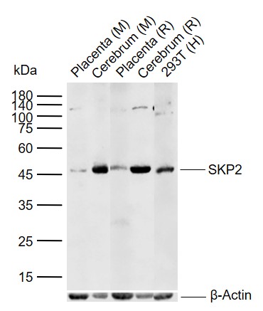 Sample:
Lane 1: Mouse Placenta tissue lysates
Lane 2: Mouse Cerebrum tissue lysates
Lane 3: Rat Placenta tissue lysates
Lane 4: Rat Cerebrum tissue lysates
Lane 5: Human 293T cell lysates
Primary: Anti-SKP2 (SL1096R) at 1/1000 dilution
Secondary: IRDye800CW Goat Anti-Rabbit IgG at 1/20000 dilution
Predicted band size: 48 kDa
Observed band size: 46 kDa
 Paraformaldehyde-fixed, paraffin embedded (mouse liver); Antigen retrieval by boiling in sodium citrate buffer (pH6.0) for 15min; Block endogenous peroxidase by 3% hydrogen peroxide for 20 minutes; Blocking buffer (normal goat serum) at 37°C for 30min; Antibody incubation with (SKP2) Polyclonal Antibody, Unconjugated (SL1096R) at 1:200 overnight at 4°C, followed by operating according to SP Kit(Rabbit) (sp-0023) instructionsand DAB staining.
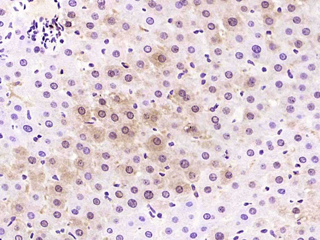 Paraformaldehyde-fixed, paraffin embedded (rat liver); Antigen retrieval by boiling in sodium citrate buffer (pH6.0) for 15min; Block endogenous peroxidase by 3% hydrogen peroxide for 20 minutes; Blocking buffer (normal goat serum) at 37°C for 30min; Antibody incubation with (SKP2) Polyclonal Antibody, Unconjugated (SL1096R) at 1:200 overnight at 4°C, followed by operating according to SP Kit(Rabbit) (sp-0023) instructionsand DAB staining.
 Tissue/cell: human thyroid gland carcinoma; 4% Paraformaldehyde-fixed and paraffin-embedded;
Antigen retrieval: citrate buffer ( 0.01M, pH 6.0 ), Boiling bathing for 15min; Block endogenous peroxidase by 3% Hydrogen peroxide for 30min; Blocking buffer (normal goat serum,C-0005) at 37℃ for 20 min;
Incubation: Anti-Skp2/skp2 p45 Polyclonal Antibody, Unconjugated(SL1096R) 1:200, overnight at 4°C, followed by conjugation to the secondary antibody(SP-0023) and DAB(C-0010) staining
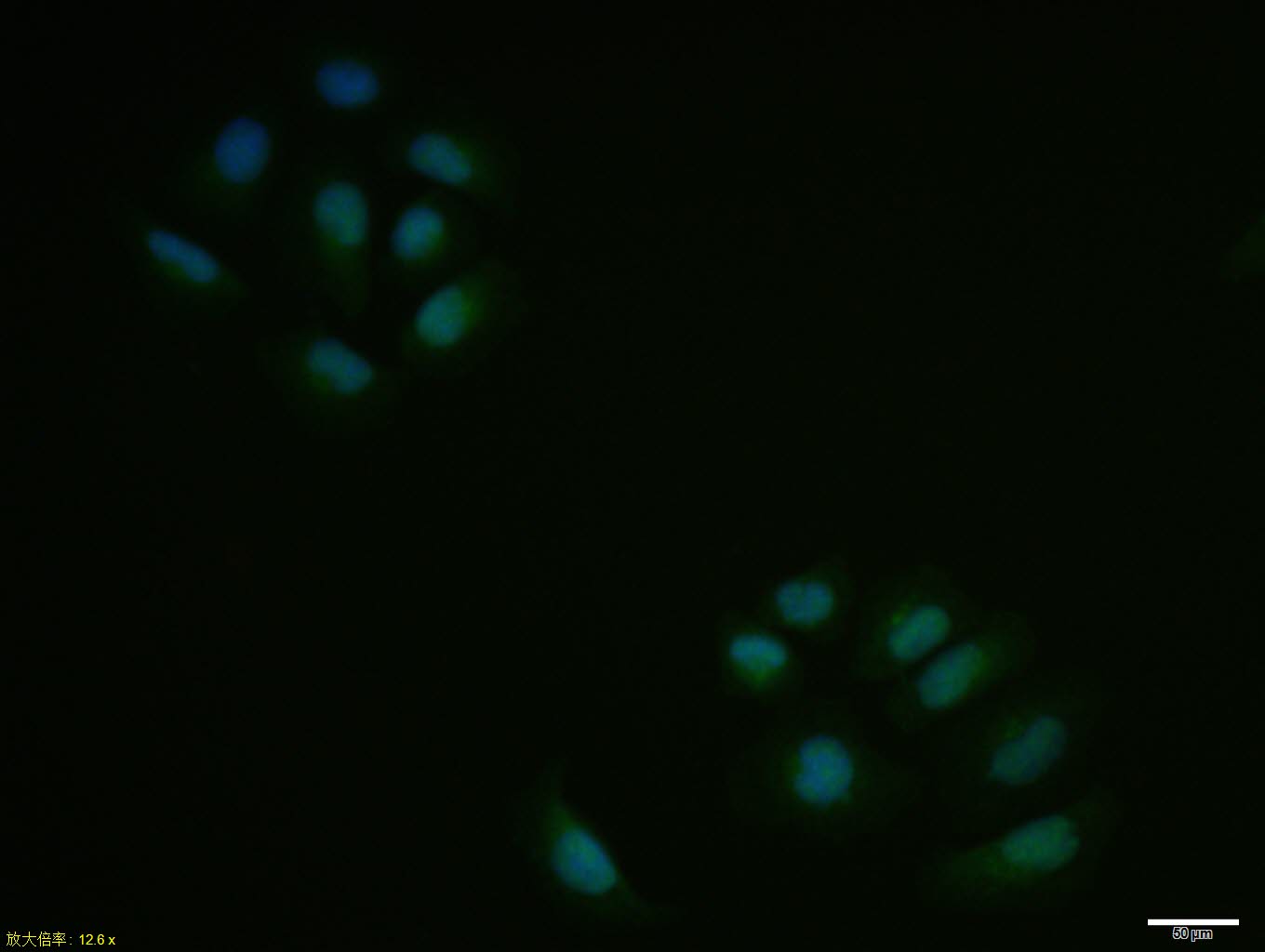 Hela cell; 4% Paraformaldehyde-fixed; Triton X-100 at room temperature for 20 min; Blocking buffer (normal goat serum, C-0005) at 37°C for 20 min; Antibody incubation with (SKP2) polyclonal Antibody, Unconjugated (SL1096R) 1:100, 90 minutes at 37°C; followed by a conjugated Goat Anti-Rabbit IgG antibody at 37°C for 90 minutes, DAPI (blue, C02-04002) was used to stain the cell nuclei.
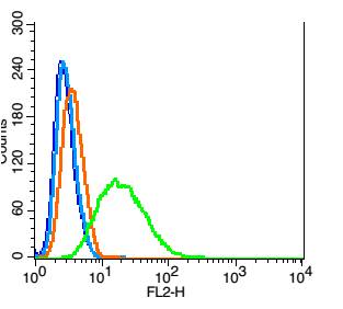 Blank control: Hela(blue).
Primary Antibody:Rabbit Anti- phospho-SKP2 antibody(SL1096R), Dilution: 1μg in 100 μL 1X PBS containing 0.5% BSA;
Isotype Control Antibody: Rabbit IgG(orange) ,used under the same conditions );
Secondary Antibody: Goat anti-rabbit IgG-PE(white blue), Dilution: 1:200 in 1 X PBS containing 0.5% BSA.
Protocol
The cells were fixed with 2% paraformaldehyde (10 min) , then permeabilized with 90% ice-cold methanol for 30 min on ice. Antibody (SL1096R, 1μg /1x10^6 cells) were incubated for 30 min on the ice, followed by 1 X PBS containing 0.5% BSA + 1 0% goat serum (15 min) to block non-specific protein-protein interactions. Then the Goat Anti-rabbit IgG/PE antibody was added into the blocking buffer mentioned above to react with the primary antibody of SL1096R at 1/200 dilution for 30 min on ice. Acquisition of 20,000 events was performed.
Cartpieces
Totalgoods,subtotals:¥Checkout
Bought notes(bought amounts latest0)
No one bought this product
User Comment(Total0User Comment Num)
- No comment


 +86 571 56623320
 +86 18668110335

