
Rabbit Anti-Laminin subunit beta-1 antibody
LAMB1; Laminin B1 chain; Laminin subunit beta 1;Laminin-1 subunit beta;Laminin-10 subunit beta;Laminin-12 subunit beta;Laminin-2 subunit beta;Laminin-6 subunit beta;Laminin-8 subunit beta;LAMB1_HUMAN.
View History [Clear]
Details
Product Name Laminin subunit beta-1 Chinese Name 层粘连蛋白β1抗体 Alias LAMB1; Laminin B1 chain; Laminin subunit beta 1; Laminin-1 subunit beta; Laminin-10 subunit beta; Laminin-12 subunit beta; Laminin-2 subunit beta; Laminin-6 subunit beta; Laminin-8 subunit beta;LAMB1_HUMAN. literatures Research Area Tumour Cell biology immunology Neurobiology Signal transduction Apoptosis Immunogen Species Rabbit Clonality Polyclonal React Species Human, Mouse, Rat, (predicted: Cow, ) Applications ELISA=1:5000-10000 IHC-P=1:100-500 IHC-F=1:100-500 IF=1:100-500 (Paraffin sections need antigen repair)
not yet tested in other applications.
optimal dilutions/concentrations should be determined by the end user.Theoretical molecular weight 198kDa Cellular localization The cell membrane Extracellular matrix Secretory protein Form Liquid Concentration 1mg/ml immunogen KLH conjugated synthetic peptide derived from human Laminin subunit beta-1: 901-1000/1786 Lsotype IgG Purification affinity purified by Protein A Buffer Solution 0.01M TBS(pH7.4) with 1% BSA, 0.03% Proclin300 and 50% Glycerol. Storage Shipped at 4℃. Store at -20 °C for one year. Avoid repeated freeze/thaw cycles. Attention This product as supplied is intended for research use only, not for use in human, therapeutic or diagnostic applications. PubMed PubMed Product Detail Laminins, a family of extracellular matrix glycoproteins, are the major noncollagenous constituent of basement membranes. They have been implicated in a wide variety of biological processes including cell adhesion, differentiation, migration, signaling, neurite outgrowth and metastasis. Laminins are composed of 3 non identical chains: laminin alpha, beta and gamma (formerly A, B1, and B2, respectively) and they form a cruciform structure consisting of 3 short arms, each formed by a different chain, and a long arm composed of all 3 chains. Each laminin chain is a multidomain protein encoded by a distinct gene. Several isoforms of each chain have been described. Different alpha, beta and gamma chain isomers combine to give rise to different heterotrimeric laminin isoforms which are designated by Arabic numerals in the order of their discovery, i.e. alpha1beta1gamma1 heterotrimer is laminin 1. The biological functions of the different chains and trimer molecules are largely unknown, but some of the chains have been shown to differ with respect to their tissue distribution, presumably reflecting diverse functions in vivo. This gene encodes the beta chain isoform laminin, beta 1. The beta 1 chain has 7 structurally distinct domains which it shares with other beta chain isomers. The C-terminal helical region containing domains I and II are separated by domain alpha, domains III and V contain several EGF-like repeats, and domains IV and VI have a globular conformation. Laminin, beta 1 is expressed in most tissues that produce basement membranes, and is one of the 3 chains constituting laminin 1, the first laminin isolated from Engelbreth-Holm-Swarm (EHS) tumor. A sequence in the beta 1 chain that is involved in cell attachment, chemotaxis, and binding to the laminin receptor was identified and shown to have the capacity to inhibit metastasis. [provided by RefSeq, Aug 2011]
Function:
Binding to cells via a high affinity receptor, laminin is thought to mediate the attachment, migration and organization of cells into tissues during embryonic development by interacting with other extracellular matrix components. Involved in the organization of the laminar architecture of cerebral cortex. It is probably required for the integrity of the basement membrane/glia limitans that serves as an anchor point for the endfeet of radial glial cells and as a physical barrier to migrating neurons. Radial glial cells play a central role in cerebral cortical development, where they act both as the proliferative unit of the cerebral cortex and a scaffold for neurons migrating toward the pial surface.
Subcellular Location:
Secreted > extracellular space > extracellular matrix > basement membrane.
Tissue Specificity:
Broadly expressed in: skin, heart, lung, and the reproductive tracts.
SWISS:
P07942
Gene ID:
3912
Database links:Entrez Gene: 10319 Human
Entrez Gene: 284217 Human
Entrez Gene: 3908 Human
Entrez Gene: 3912 Human
Entrez Gene: 3913 Human
Entrez Gene: 3915 Human
Entrez Gene: 3918 Human
Entrez Gene: 16772 Mouse
Omim: 150320 Human
SwissProt: P07942 Human
SwissProt: P24043 Human
SwissProt: P25391 Human
SwissProt: P55268 Human
SwissProt: Q9Y6N6 Human
SwissProt: P19137 Mouse
SwissProt: Q9R0B6 Mouse
Unigene: 201805 Human
Unigene: 270364 Human
Unigene: 302362 Mouse
层粘连蛋白主要存在于基膜(basal lamina)结构中,是基膜所特有的非胶原glycoprotein。
层粘连蛋白在细胞发育过程中刺激细胞粘着、细胞运动。LN能够刺激胚胎中神经轴的生长,并促进成年动物的神经损伤后重生长和再生。如同纤粘连蛋白,细胞外的LN能够影响细胞的生长、迁移和分化。LN在原生殖细胞的迁移中起关键作用。
层粘连蛋白是细胞外基底膜的主要组成部分,分布于基底膜和Extracellular matrix内、成纤维细胞、endothelial cells和平滑肌细胞分泌层粘连蛋白。Tumour的浸润和转移与其遭到破坏相关。该抗体可用于标记基底膜,有助于观察Tumour周围基底膜的分布及其改变,间接提示该Tumour的分化程度和浸润情况。Product Picture 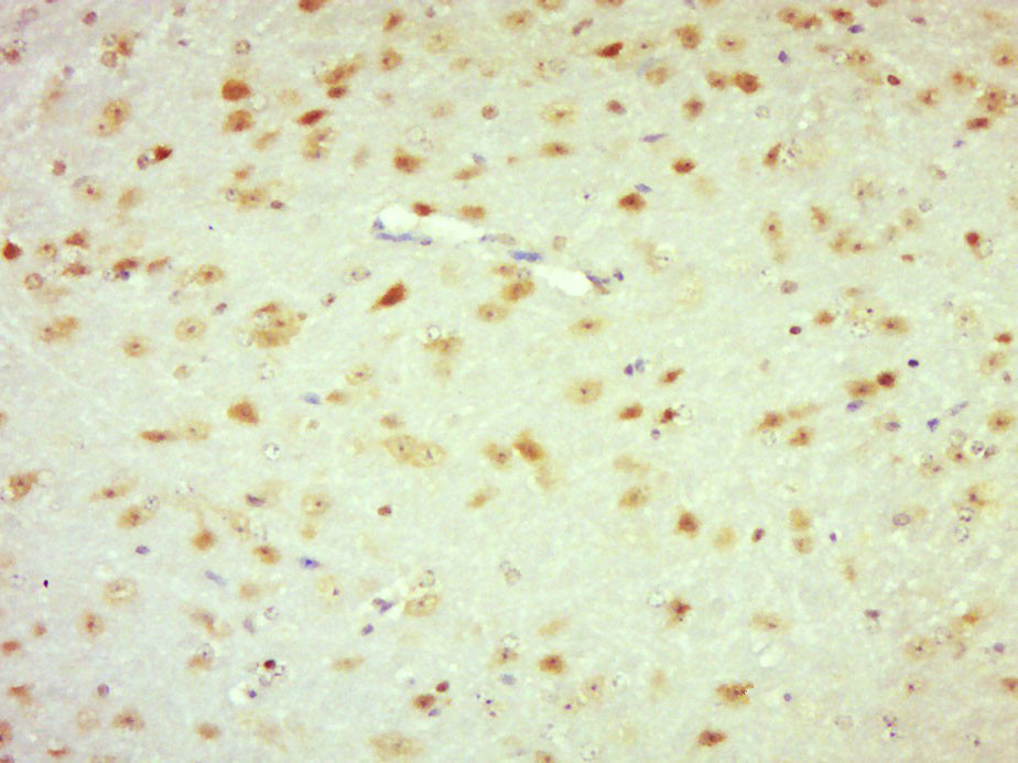 Paraformaldehyde-fixed, paraffin embedded (Mouse brain); Antigen retrieval by boiling in sodium citrate buffer (pH6.0) for 15min; Block endogenous peroxidase by 3% hydrogen peroxide for 20 minutes; Blocking buffer (normal goat serum) at 37°C for 30min; Antibody incubation with (laminin) Polyclonal Antibody, Unconjugated (SL0821R) at 1:500 overnight at 4°C, followed by a conjugated secondary (sp-0023) for 20 minutes and DAB staining.
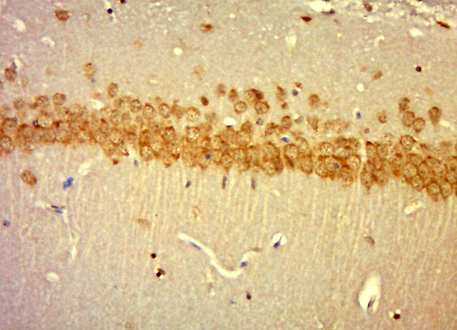 Paraformaldehyde-fixed, paraffin embedded (mouse brain); Antigen retrieval by boiling in sodium citrate buffer (pH6.0) for 15min; Block endogenous peroxidase by 3% hydrogen peroxide for 20 minutes; Blocking buffer (normal goat serum) at 37°C for 30min; Antibody incubation with (laminin) Polyclonal Antibody, Unconjugated (SL0821R) at 1:400 overnight at 4°C, followed by a conjugated secondary (sp-0023) for 20 minutes and DAB staining.
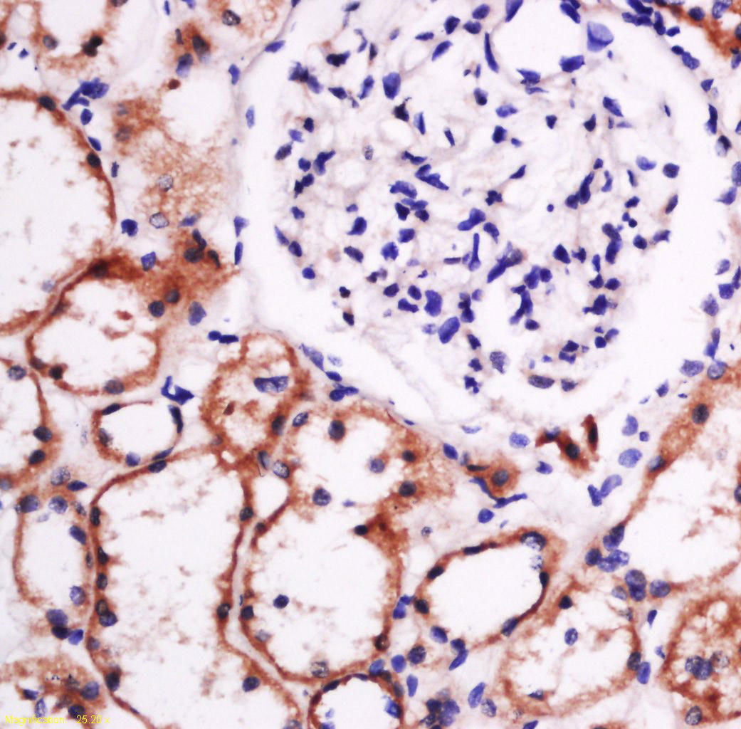 Paraformaldehyde-fixed, paraffin embedded (human kidney tissue); Antigen retrieval by boiling in sodium citrate buffer (pH6.0) for 15min; Block endogenous peroxidase by 3% hydrogen peroxide for 20 minutes; Blocking buffer (normal goat serum) at 37°C for 30min; Antibody incubation with (laminin) Polyclonal Antibody, Unconjugated (SL0821R) at 1:400 overnight at 4°C, followed by a conjugated secondary (sp-0023) for 20 minutes and DAB staining.
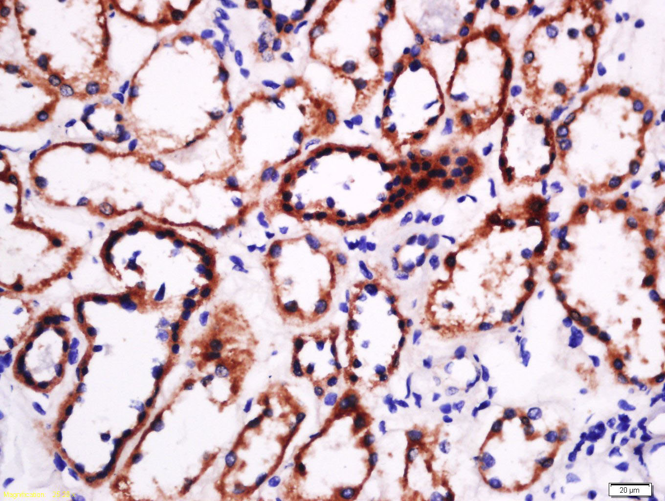 Tissue/cell: human kidney tissue; 4% Paraformaldehyde-fixed and paraffin-embedded;
Antigen retrieval: citrate buffer ( 0.01M, pH 6.0 ), Boiling bathing for 15min; Block endogenous peroxidase by 3% Hydrogen peroxide for 30min; Blocking buffer (normal goat serum,C-0005) at 37℃ for 20 min;
Incubation: Anti-laminin Polyclonal Antibody, Unconjugated(SL0821R) 1:500, overnight at 4°C, followed by conjugation to the secondary antibody(SP-0023) and DAB(C-0010) staining
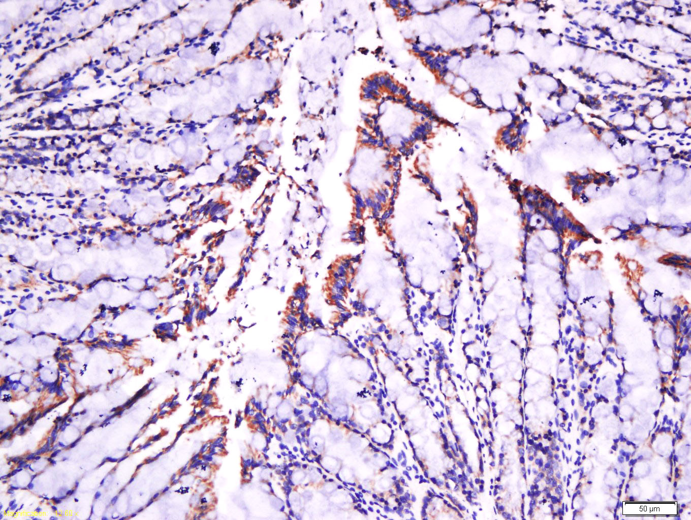 Tissue/cell: rat intestine tissue; 4% Paraformaldehyde-fixed and paraffin-embedded;
Antigen retrieval: citrate buffer ( 0.01M, pH 6.0 ), Boiling bathing for 15min; Block endogenous peroxidase by 3% Hydrogen peroxide for 30min; Blocking buffer (normal goat serum,C-0005) at 37℃ for 20 min;
Incubation: Anti-laminin Polyclonal Antibody, Unconjugated(SL0821R) 1:500, overnight at 4°C, followed by conjugation to the secondary antibody(SP-0023) and DAB(C-0010) staining
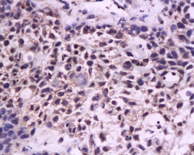 Tissue/cell: Transplantation tumor of In nude mice; 4% Paraformaldehyde-fixed and paraffin-embedded;
Antigen retrieval: citrate buffer ( 0.01M, pH 6.0 ), Boiling bathing for 15min; Block endogenous peroxidase by 3% Hydrogen peroxide for 30min; Blocking buffer (normal goat serum,C-0005) at 37℃ for 20 min;
Incubation: Anti-laminin Polyclonal Antibody, Unconjugated(SL0821R) 1:200, overnight at 4°C, followed by conjugation to the secondary antibody(SP-0023) and DAB(C-0010) staining
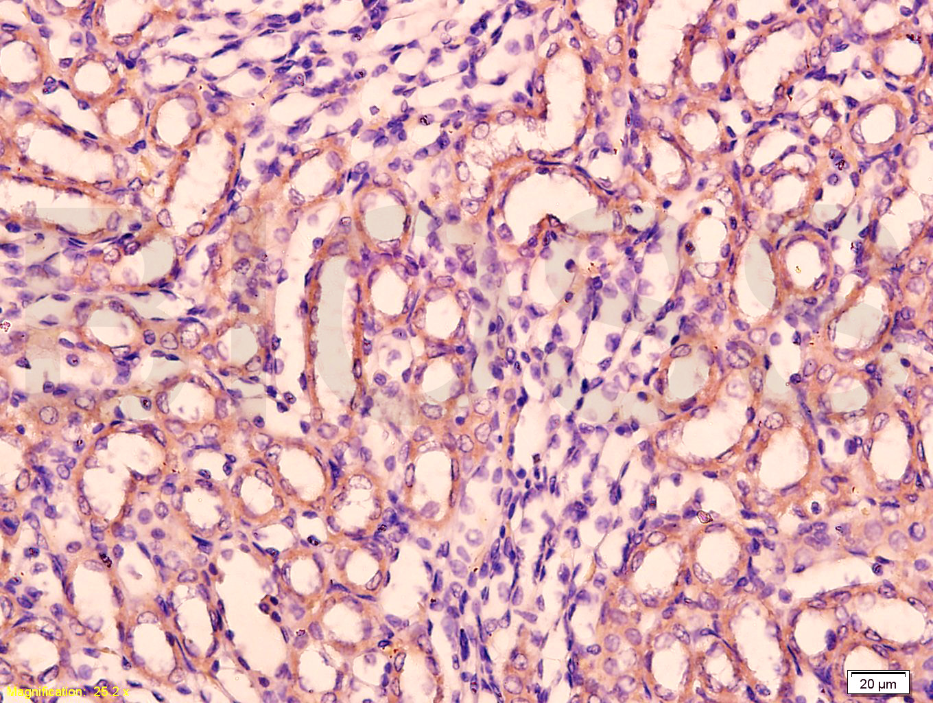 Tissue/cell: rat kidney tissue; 4% Paraformaldehyde-fixed and paraffin-embedded;
Antigen retrieval: citrate buffer ( 0.01M, pH 6.0 ), Boiling bathing for 15min; Block endogenous peroxidase by 3% Hydrogen peroxide for 30min; Blocking buffer (normal goat serum,C-0005) at 37℃ for 20 min;
Incubation: Anti-laminin Polyclonal Antibody, Unconjugated(SL0821R) 1:200, overnight at 4°C, followed by conjugation to the secondary antibody(SP-0023) and DAB(C-0010) staining
Cartpieces
Totalgoods,subtotals:¥Checkout
Bought notes(bought amounts latest0)
No one bought this product
User Comment(Total0User Comment Num)
- No comment


 +86 571 56623320
 +86 18668110335

