
Rabbit Anti-CD4 antibody
CD4 (L3T4); CD4 antigen (p55); CD4 Antigen ; CD4 molecule; CD4 Receptor; CD4+ Lymphocyte deficiency, included; CD4mut; L3T4; Leu3; Ly-4; Lymphocyte antigen CD4; MGC165891; p55; T Cell Antigen T4 ; T cell antigen T4/LEU3; T cell differentiation antigen L3T
View History [Clear]
Details
Product Name CD4 Chinese Name CD4抗体 Alias CD4 (L3T4); CD4 antigen (p55); CD4 Antigen ; CD4 molecule; CD4 Receptor; CD4+ Lymphocyte deficiency, included; CD4mut; L3T4; Leu3; Ly-4; Lymphocyte antigen CD4; MGC165891; p55; T Cell Antigen T4 ; T cell antigen T4/LEU3; T cell differentiation antigen L3T4; T cell OKT4 deficiency, included; T cell surface antigen T4/Leu 3 ; T cell surface antigen T4/Leu3; T Cell Surface Glycoprotein CD4; W3/25; W3/25 antigen; T-cell surface glycoprotein CD4 isoform 1 precursor; CD4_MOUSE. literatures Research Area Cell biology immunology Stem cells Cell Surface Molecule lymphocyte t-lymphocyte Immunogen Species Rabbit Clonality Polyclonal React Species Human, Mouse, Rat, Applications WB=1:500-2000 ELISA=1:5000-10000 IHC-P=1:100-500 IHC-F=1:100-500 IF=1:100-500 (Paraffin sections need antigen repair)
not yet tested in other applications.
optimal dilutions/concentrations should be determined by the end user.Theoretical molecular weight 48kDa Detection molecular weight 55 kDa Cellular localization The cell membrane Form Liquid Concentration 1mg/ml immunogen KLH conjugated synthetic peptide derived from the middle of mouse CD4: 231-330/457 <Extracellular> Lsotype IgG Purification affinity purified by Protein A Buffer Solution 0.01M TBS(pH7.4) with 1% BSA, 0.03% Proclin300 and 50% Glycerol. Storage Shipped at 4℃. Store at -20 °C for one year. Avoid repeated freeze/thaw cycles. Attention This product as supplied is intended for research use only, not for use in human, therapeutic or diagnostic applications. PubMed PubMed Product Detail This gene encodes the CD4 membrane glycoprotein of T lymphocytes. The CD4 antigen acts as a coreceptor with the T-cell receptor on the T lymphocyte to recognize antigens displayed by an antigen presenting cell in the context of class II MHC molecules. The CD4 antigen is also a primary receptor for entry of the human immunodeficiency virus through interactions with the HIV Env gp120 subunit. This gene is expressed not only in T lymphocytes, but also in B cells, macrophages, granulocytes, as well as in various regions of the brain. The protein functions to initiate or augment the early phase of T-cell activation, and may function as an important mediator of indirect neuronal damage in infectious and immune-mediated diseases of the central nervous system. Multiple alternatively spliced transcript variants encoding different isoforms have been identified in this gene. [provided by RefSeq, May 2020]
Function:
Accessory protein for MHC class-II antigen/T-cell receptor interaction. May regulate T-cell activation. Induces the aggregation of lipid rafts.
Subunit:
Associates with LCK. Binds to HIV-1 gp120 and to P4HB/PDI and upon HIV-1 binding to the cell membrane, is part of P4HB/PDI-CD4-CXCR4-gp120 complex. Interacts with HIV-1 Envelope polyprotein gp160 and protein Vpu. Interacts with Human Herpes virus 7 capsid proteins. Interacts with PTK2/FAK1; this interaction requires the presence of HIV-1 gp120.
Subcellular Location:
Cell membrane; Single-pass type I membrane protein. Note=Localizes to lipid rafts. Removed from plasma membrane by HIV-1 Nef protein that increases clathrin-dependent endocytosis of this antigen to target it to lysosomal degradation. Cell surface expression is also down-modulated by HIV-1 Envelope polyprotein gp160 that interacts with, and sequesters CD4 in the endoplasmic reticulum.
Post-translational modifications:
Palmitoylation and association with LCK contribute to the enrichment of CD4 in lipid rafts.
Similarity:
Contains 3 Ig-like C2-type (immunoglobulin-like) domains.
Contains 1 Ig-like V-type (immunoglobulin-like) domain.
SWISS:
P06332
Gene ID:
12504
Database links:
Entrez Gene: 920 Human
Entrez Gene: 12504 Mouse
Omim: 186940 Human
SwissProt: P06332 Mouse
SwissProt: P01730 Human
Unigene: 631659 Human
Unigene: 2209 Mouse
此抗体可识别55KDⅠ型单链穿膜glycoprotein。
CD4分子是存在于大多数辅助/诱导T细胞表面的59kDa的glycoprotein。正常淋巴组织中CD4的表达数量多于CD8,此抗体主要用于标记辅助/诱导T细胞,与CD8单抗联合使用对外周血lymphocyte分型。
CD4抗原是HLA-II类分子和人类免疫缺陷病毒(HIV)-爱滋病的受体,在35-50%外周血lymphocyte-辅助和诱导T细胞(Th/Ti)和70-80%人胸腺细胞上表达,在人的单核细胞表面也有低密度的表达。
CD4抗原有膜结合型和可溶性两种形式。Th/Ti可辅助Ig产生和T细胞毒T细胞的作用。Product Picture 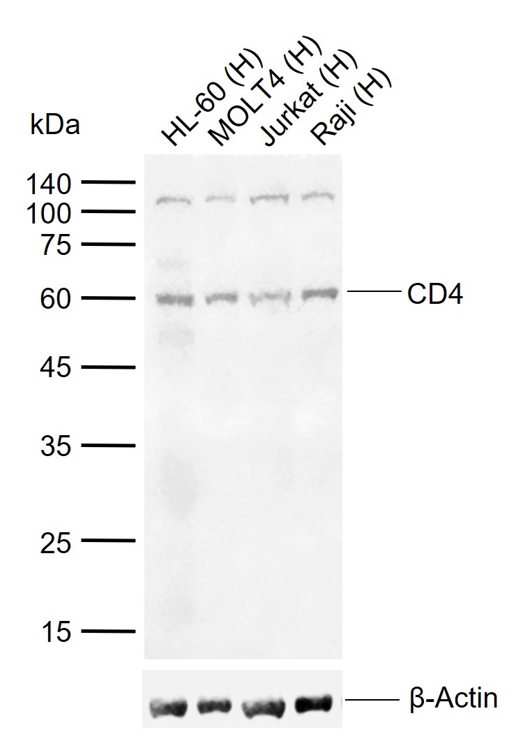 Sample:
Lane 1: Human HL-60 cell lysates
Lane 2: Human MOLT4 cell lysates
Lane 3: Human Jurkat cell lysates
Lane 4: Human Raji cell lysates
Primary: Anti-CD4 (SL0766R) at 1/1000 dilution
Secondary: IRDye800CW Goat Anti-Rabbit IgG at 1/20000 dilution
Predicted band size: 48 kDa
> Observed band size: 60 kDa
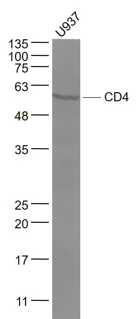 Sample:
U937(Human) Cell Lysate at 30 ug
Primary: Anti- CD4 (SL0766R) at 1/1000 dilution
Secondary: IRDye800CW Goat Anti-Rabbit IgG at 1/20000 dilution
Predicted band size: 48 kD
Observed band size: 55 kD
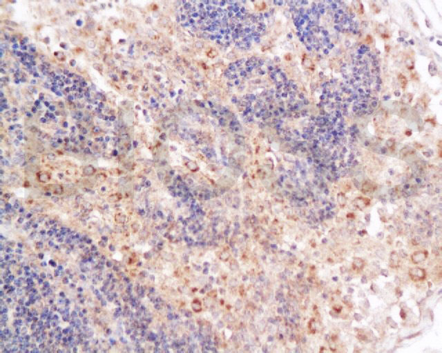
 Paraformaldehyde-fixed, paraffin embedded (rat spleen); Antigen retrieval by boiling in sodium citrate buffer (pH6.0) for 15min; Block endogenous peroxidase by 3% hydrogen peroxide for 20 minutes; Blocking buffer (normal goat serum) at 37°C for 30min; Antibody incubation with (CD4) Polyclonal Antibody, Unconjugated (SL0766R) at 1:400 overnight at 4°C, followed by a conjugated secondary (sp-0023) for 20 minutes and DAB staining.
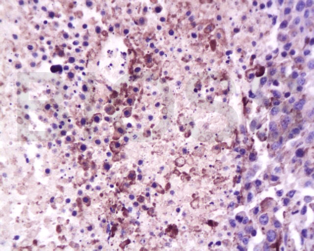 Tissue/cell: mouse lymphoma tissue; 4% Paraformaldehyde-fixed and paraffin-embedded;
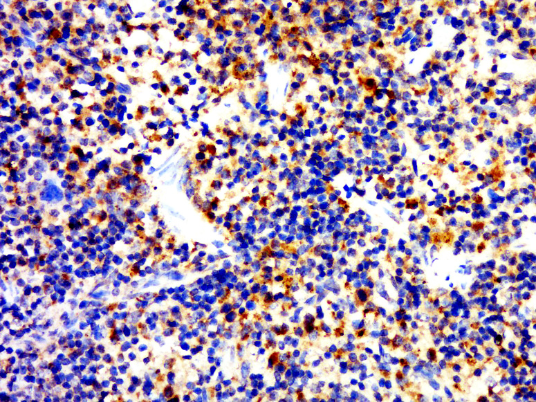
Antigen retrieval: citrate buffer ( 0.01M, pH 6.0 ), Boiling bathing for 15min; Block endogenous peroxidase by 3% Hydrogen peroxide for 30min; Blocking buffer (normal goat serum,C-0005) at 37℃ for 20 min;
Incubation: Anti-CD4 Polyclonal Antibody, Unconjugated(SL0766R) 1:200, overnight at 4癈, followed by conjugation to the secondary antibody(SP-0023) and DAB(C-0010) staining
 Tissue/cell: mouse lymphoma tissue; 4% Paraformaldehyde-fixed and paraffin-embedded;
Antigen retrieval: citrate buffer ( 0.01M, pH 6.0 ), Boiling bathing for 15min; Block endogenous peroxidase by 3% Hydrogen peroxide for 30min; Blocking buffer (normal goat serum,C-0005) at 37℃ for 20 min;
Incubation: Anti-CD4 Polyclonal Antibody, Unconjugated(SL0766R) 1:200, overnight at 4癈, followed by conjugation to the secondary antibody(SP-0023) and DAB(C-0010) staining
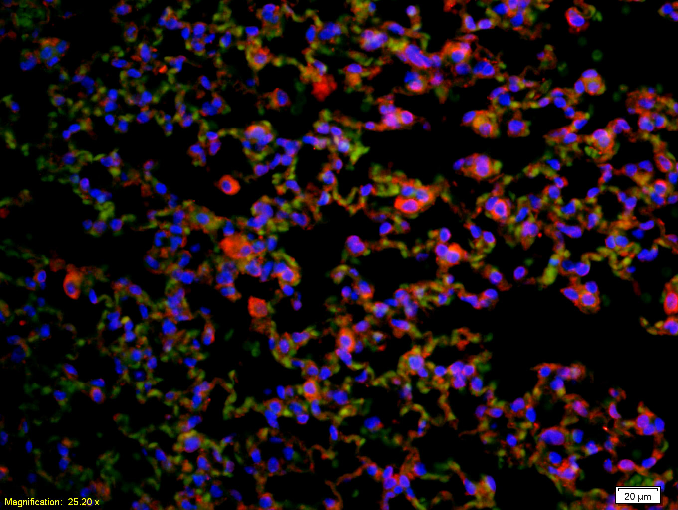 Tissue/cell: rat lung tissue;4% Paraformaldehyde-fixed and paraffin-embedded;
Antigen retrieval: citrate buffer ( 0.01M, pH 6.0 ), Boiling bathing for 15min; Blocking buffer (normal goat serum,C-0005) at 37℃ for 20 min;
Incubation: Anti-CD4(mouse, rat) Polyclonal Antibody, Unconjugated(SL0766R) 1:200, overnight at 4癈; The secondary antibody was Goat Anti-Rabbit IgG, Cy3 conjugated(SL0295G-Cy3)used at 1:200 dilution for 40 minutes at 37癈. DAPI(5ug/ml,blue,C-0033) was used to stain the cell nuclei
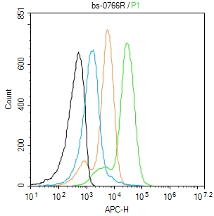 Blank control: Mouse spleen.
Primary Antibody (green line): Rabbit Anti-CD4 antibody (SL0766R)
Dilution: 3μg /10^6 cells;
Isotype Control Antibody (orange line): Rabbit IgG .
Secondary Antibody: Goat anti-rabbit IgG-AF647
Dilution: 3μg /test.
Protocol
The cells incubated in 5%BSA to block non-specific protein-protein interactions for 30 min at room temperature .Cells stained with Primary Antibody for 30 min at room temperature. The secondary antibody used for 40 min at room temperature. Acquisition of 20,000 events was performed.Blank control: Mouse spleen.
Primary Antibody (green line): Rabbit Anti-CD4 antibody (SL0766R)
Dilution: 3μg /10^6 cells;
Isotype Control Antibody (orange line): Rabbit IgG .
Secondary Antibody: Goat anti-rabbit IgG-AF647
Dilution: 3μg /test.
Protocol
The cells incubated in 5%BSA to block non-specific protein-protein interactions for 30 min at room temperature .Cells stained with Primary Antibody for 30 min at room temperature. The secondary antibody used for 40 min at room temperature. Acquisition of 20,000 events was performed.
Cartpieces
Totalgoods,subtotals:¥Checkout
Bought notes(bought amounts latest0)
No one bought this product
User Comment(Total0User Comment Num)
- No comment


 +86 571 56623320
 +86 18668110335

