
Rabbit Anti-CDKN1B/p27 KIP 1 antibody
p27 KIP 1; CDN1B_HUMAN; AA408329; AI843786; Cdki1b; CDKN 1B; CDKN 4; CDKN1B; CDKN1B; CDKN4; CDKN4; Cyclin Dependent Kinase Inhibitor 1B; Cyclin Dependent Kinase Inhibitor 1B; Cyclin dependent kinase inhibitor p27; Cyclin dependent kinase inhibitor p27; Cy
View History [Clear]
Details
Product Name CDKN1B/p27 KIP 1 Chinese Name P27抗体/周期素依赖激酶抑制剂 Alias p27 KIP 1; CDN1B_HUMAN; AA408329; AI843786; Cdki1b; CDKN 1B; CDKN 4; CDKN1B; CDKN1B; CDKN4; CDKN4; Cyclin Dependent Kinase Inhibitor 1B; Cyclin Dependent Kinase Inhibitor 1B; Cyclin dependent kinase inhibitor p27; Cyclin dependent kinase inhibitor p27; Cyclin-dependent kinase inhibitor 1B (p27, Kip1); Cyclin-dependent kinase inhibitor 1B; Cyclin-dependent kinase inhibitor p27; Cyclin-dependent kinase inhibitor p27 Kip1; KIP 1; KIP1; MEN1B; MEN4; OTTHUMP00000195098; OTTHUMP00000195099; p27; p27 Kip1; P27-like cyclin-dependent kinase inhibitor; P27KIP1. literatures Research Area Tumour immunology Chromatin and nuclear signals Signal transduction Apoptosis Immunogen Species Rabbit Clonality Polyclonal React Species Human, Mouse, Rat, (predicted: Chicken, Dog, Pig, Sheep, ) Applications WB=1:500-2000 ELISA=1:5000-10000 IP=1:20-100 IHC-P=1:100-500 IHC-F=1:100-500 Flow-Cyt=1μg/Test IF=1:200-800 (Paraffin sections need antigen repair)
not yet tested in other applications.
optimal dilutions/concentrations should be determined by the end user.Theoretical molecular weight 22kDa Cellular localization The nucleus cytoplasmic Form Liquid Concentration 1mg/ml immunogen KLH conjugated synthetic peptide derived from human P27 kip1: 101-198/198 Lsotype IgG Purification affinity purified by Protein A Buffer Solution 0.01M TBS(pH7.4) with 1% BSA, 0.03% Proclin300 and 50% Glycerol. Storage Shipped at 4℃. Store at -20 °C for one year. Avoid repeated freeze/thaw cycles. Attention This product as supplied is intended for research use only, not for use in human, therapeutic or diagnostic applications. PubMed PubMed Product Detail Cell cycle progression is regulated by cyclins and their cognate Cdks. p27 KIP 1 is a cell cycle regulatory mitotic inhibitor of cdk activity. p27 KIP 1 is a candidate tumor suppressor gene, and has been proposed to function as a possible mediator of TGF beta induced G1 arrest. p27 KIP 1 is up regulated in response to antimitogenic stimuli. The increased protein expression of p27 results in cellular arrest by binding to cyclin/Cdk complexes such as cyclin D1/Cdk4. p27 Kip1 is regulated by phosphorylation on serine 10 (S10) and threonine 187 (T187). Phosphorylation by CDK2 on T187 results in ubiquitylation and degradation of p27 Kip 1; while phosphorylation by hKIS on S10 signals the nuclear export to the cytoplasm.
Function:
Important regulator of cell cycle progression. Involved in G1 arrest. Potent inhibitor of cyclin E- and cyclin A-CDK2 complexes. Forms a complex with cyclin type D-CDK4 complexes and is involved in the assembly, stability, and modulation of CCND1-CDK4 complex activation. Acts either as an inhibitor or an activator of cyclin type D-CDK4 complexes depending on its phosphorylation state and/or stoichometry.
Subunit:
Forms a ternary compex with CCNE1/CDK2/CDKN1B.
Subcellular Location:
Nucleus. Cytoplasm. Endosome. Note=Nuclear and cytoplasmic in quiescent cells. AKT-or RSK-mediated phosphorylation on Thr-198, binds 14-3-3, translocates to the cytoplasm and promotes cell cycle progression. Mitogen-activated UHMK1 phosphorylation on Ser-10 also results in translocation to the cytoplasm and cell cycle progression. Phosphorylation on Ser-10 facilitates nuclear export. Translocates to the nucleus on phosphorylation of Tyr-88 and Tyr-89. Colocalizes at the endosome with SNX6; this leads to lysosomal degradation.
Tissue Specificity:
Expressed in all tissues tested. Highest levels in skeletal muscle, lowest in liver and kidney.
Post-translational modifications:
Phosphorylated; phosphorylation occurs on serine, threonine and tyrosine residues. Phosphorylation on Ser-10 is the major site of phosphorylation in resting cells, takes place at the G(0)-G(1) phase and leads to protein stability. Phosphorylation on other sites is greatly enhanced by mitogens, growth factors, cMYC and in certain cancer cell lines. The phosphorylated form found in the cytoplasm is inactivate. Phosphorylation on Thr-198 is required for interaction with 14-3-3 proteins. Phosphorylation on Thr-187, by CDK2 leads to protein ubiquitination and proteasomal degradation. Tyrosine phosphorylation promotes this process. Phosphorylation by PKB/AKT1 can be suppressed by LY294002, an inhibitor of the catalytic subunit of PI3K. Phosphorylation on Tyr-88 and Tyr-89 has no effect on binding CDK2, but is required for binding CDK4. Dephosphorylated on tyrosine residues by G-CSF.
Ubiquitinated; in the cytoplasm by the KPC complex (composed of RNF123/KPC1 and UBAC1/KPC2) and, in the nucleus, by SCF(SKP2). The latter requires prior phosphorylation on Thr-187. Ubiquitinated; by a TRIM21-containing SCF(SKP2)-like complex; leads to its degradation.
DISEASE:
Defects in CDKN1B are the cause of multiple endocrine neoplasia type 4 (MEN4) [MIM:610755]. Multiple endocrine neoplasia (MEN) syndromes are inherited cancer syndromes of the thyroid. MEN4 is a MEN-like syndrome with a phenotypic overlap of both MEN1 and MEN2.
Similarity:
Belongs to the CDI family.
SWISS:
P46527
Gene ID:
1027
Database links:Entrez Gene: 1027 Human
Entrez Gene: 12576 Mouse
Omim: 600778 Human
SwissProt: P46527 Human
SwissProt: P46414 Mouse
Unigene: 238990 Human
Unigene: 2958 Mouse
P27蛋白是一种新发现的周期素依赖激酶抑制剂,属于细胞周期的负性调控因子。P27基因及其产物的异常表达可能与某些Tumour的发生、发展有着密切的关系。P27蛋白对细胞周期的调控及在Tumour中发挥着很重要的作用,The nucleus表达。Product Picture 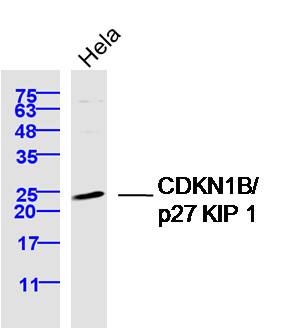 Sample: Hela Cell (Human) Lysate at 40 ug
Primary: Anti-CDKN1B/p27 KIP 1 (SL0742R) at 1/300 dilution
Secondary: IRDye800CW Goat Anti-Rabbit IgG at 1/20000 dilution
Predicted band size: 22 kD
Observed band size: 24 kD
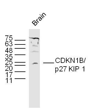 Sample:
Brain (Mouse) Lysate at 40 ug
Primary: Anti- CDKN1B'p27 KIP 1 (SL0742R) at 1/300 dilution
Secondary: IRDye800CW Goat Anti-Rabbit IgG at 1/20000 dilution
Predicted band size: 22 kD
Observed band size: 22 kD
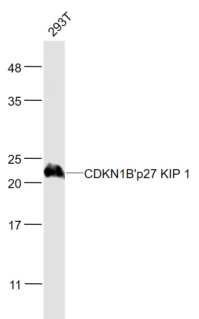 Sample:
293T(Human) Cell Lysate at 30 ug
Primary: Anti- CDKN1B'p27 KIP 1 (SL0742R) at 1/1000 dilution
Secondary: IRDye800CW Goat Anti-Rabbit IgG at 1/20000 dilution
Predicted band size: 22 kD
Observed band size: 22 kD
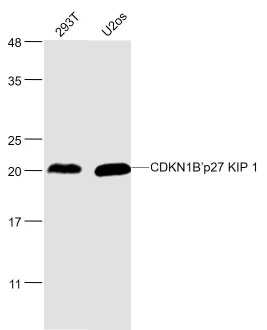 Sample:
293T(Human) Cell Lysate at 30 ug
U2os(Human) Cell Lysate at 30 ug
Primary: Anti- CDKN1B’p27 KIP 1 (SL0742R) at 1/1000 dilution
Secondary: IRDye800CW Goat Anti-Rabbit IgG at 1/20000 dilution
Predicted band size: 22 kD
Observed band size: 22 kD
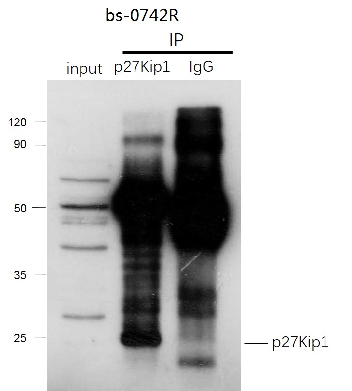 CDKN1B (p27Kip1) was immunoprecipitated from mouse kidney tissue with SL0742R at 1/150 dilution. Western blot was performed from the immunoprecipitate using protein A/G beads. HRP Conjugated Goat anti-Rabbit IgG (Heavy Chain specific) was used as secondary antibody at 1:5000 dilution.
Lane 1: mouse kidney tissue lysate 10 µg (Input).
Lane 2: SL0742R IP in mouse kidney tissue lysate.
Lane 3: native rabbit IgG IP in mouse kidney tissue lysate (negative control).
Secondary
All lanes Goat anti-Rabbit IgG (Heavy Chain specific), HRP Conjugated, 1:5000
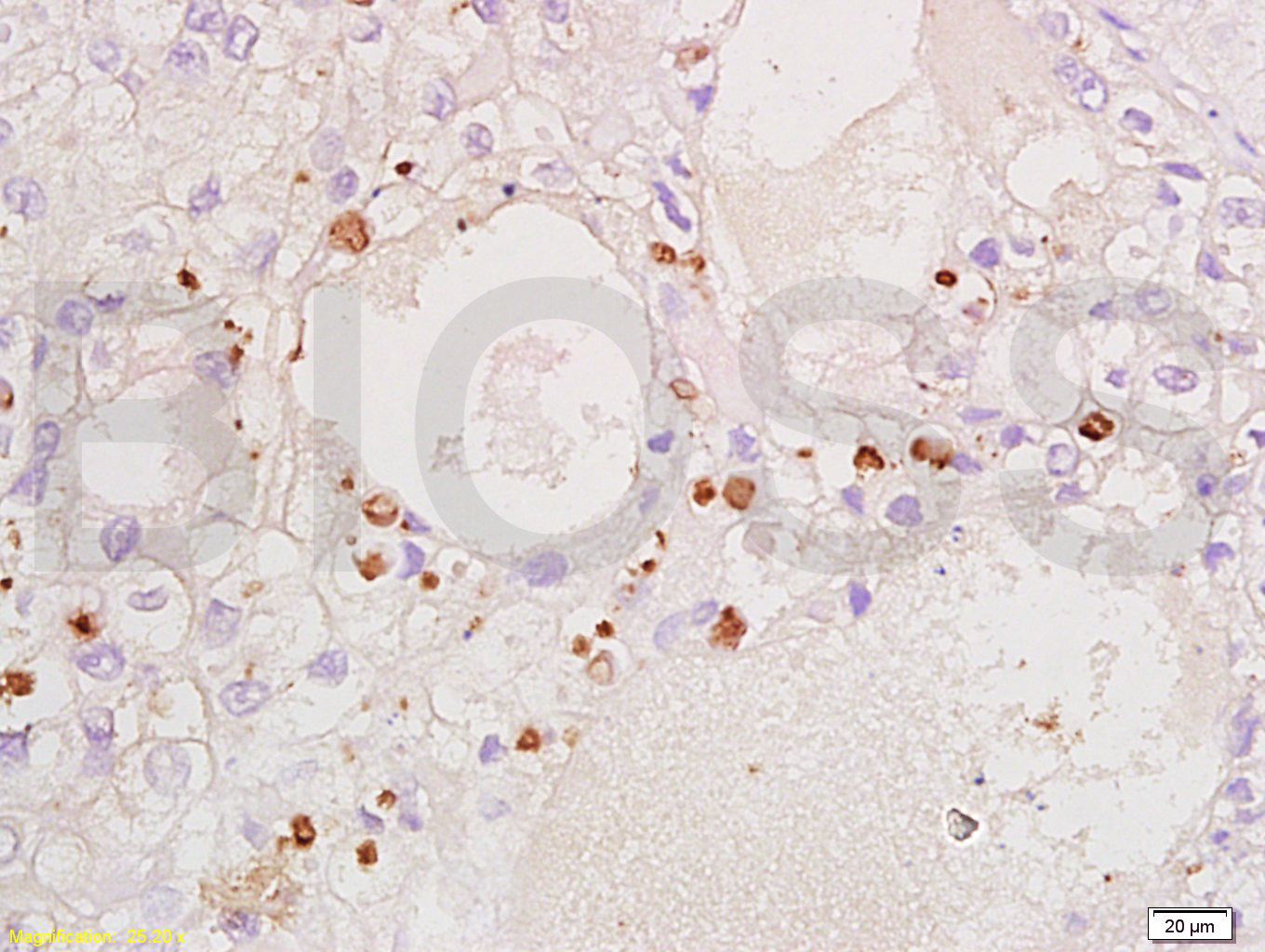 Tissue/cell: human ovary carcinoma; 4% Paraformaldehyde-fixed and paraffin-embedded;
Antigen retrieval: citrate buffer ( 0.01M, pH 6.0 ), Boiling bathing for 15min; Block endogenous peroxidase by 3% Hydrogen peroxide for 30min; Blocking buffer (normal goat serum,C-0005) at 37∩ for 20 min;
Incubation: Anti-CDKN1B/P27kip1 Polyclonal Antibody, Unconjugated(SL0742R) 1:200, overnight at 4∑C, followed by conjugation to the secondary antibody(SP-0023) and DAB(C-0010) staining
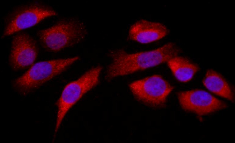 Tissue/cell: MCF7; 4% Paraformaldehyde-fixed; Triton X-100 at room temperature for 20 min; Blocking buffer (normal goat serum, C-0005) at 37°C for 20 min; Antibody incubation with (CDKN1B ) Polyclonal Antibody, Unconjugated (SL0742R) 1:200, 90 minutes at 37°C; followed by a conjugated Goat Anti-Rabbit IgG antibody (SL0295G-FITC) at 37°C for 90 minutes, DAPI (blue, C02-04002) was used to stain the cell nuclei.
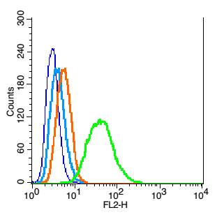 Blank control: RSC96(blue), the cells were fixed with 2% paraformaldehyde (10 min) and then permeabilized with ice-cold 90% methanol for 30 min on ice.
Isotype Control Antibody: Rabbit IgG(orange) ; Secondary Antibody: Goat anti-rabbit IgG-PE(white blue), Dilution: 1:200 in 1 X PBS containing 0.5% BSA ; Primary Antibody Dilution: 1μg in 100μL1X PBS containing 0.5% BSA(green).
Cartpieces
Totalgoods,subtotals:¥Checkout
Bought notes(bought amounts latest0)
No one bought this product
User Comment(Total0User Comment Num)
- No comment


 +86 571 56623320
 +86 18668110335

