
Rabbit Anti-Collagen II antibody
Collagen II alpha 1; COL2A1; COL2A1 protein; collagen, type II, alpha 1; collagen alpha-1(II); type II collagen; alpha-1 type II collagen; alpha1 type II collagen; Col2a1; AOM; Cartilage collagen; Chondrocalcin; COL11A3; Collagen alpha 1(II) chain precur
View History [Clear]
Details
Product Name Collagen II Chinese Name Ⅱ型胶原α1蛋白/软骨钙素抗体 Alias Collagen II alpha 1; COL2A1; COL2A1 protein; collagen, type II, alpha 1; collagen alpha-1(II); type II collagen; alpha-1 type II collagen; alpha1 type II collagen; Col2a1; AOM; Cartilage collagen; Chondrocalcin; COL11A3; Collagen alpha 1(II) chain precursor; Collagen II alpha 1 polypeptide; Collagen type II alpha 1 (primary osteoarthritis spondyloepiphyseal dysplasia congenital); MGC131516; SEDC; Collagen alpha-1(II) chain; Alpha-1 type II collagen; CO2A1_HUMAN. literatures Research Area Tumour Cell biology immunology Immunogen Species Rabbit Clonality Polyclonal React Species Human, Rabbit, (predicted: Mouse, Rat, Chicken, Dog, Pig, Cow, Guinea Pig, ) Applications WB=1:500-2000 ELISA=1:5000-10000 IHC-P=1:100-500 IHC-F=1:100-500 IF=1:100-500 (Paraffin sections need antigen repair)
not yet tested in other applications.
optimal dilutions/concentrations should be determined by the end user.Theoretical molecular weight 117kDa Cellular localization Extracellular matrix Secretory protein Form Liquid Concentration 1mg/ml immunogen KLH conjugated synthetic peptide derived from human Collagen II: 1201-1300/1487 Lsotype IgG Purification affinity purified by Protein A Buffer Solution 0.01M TBS(pH7.4) with 1% BSA, 0.03% Proclin300 and 50% Glycerol. Storage Shipped at 4℃. Store at -20 °C for one year. Avoid repeated freeze/thaw cycles. Attention This product as supplied is intended for research use only, not for use in human, therapeutic or diagnostic applications. PubMed PubMed Product Detail This gene encodes the alpha-1 chain of type II collagen, a fibrillar collagen found in cartilage and the vitreous humor of the eye. Mutations in this gene are associated with achondrogenesis, chondrodysplasia, early onset familial osteoarthritis, SED congenita, Langer-Saldino achondrogenesis, Kniest dysplasia, Stickler syndrome type I, and spondyloepimetaphyseal dysplasia Strudwick type. In addition, defects in processing chondrocalcin, a calcium binding protein that is the C-propeptide of this collagen molecule, are also associated with chondrodysplasia. There are two transcripts identified for this gene. [provided by RefSeq, Jul 2008]
Function:
Type II collagen is specific for cartilaginous tissues. It is essential for the normal embryonic development of the skeleton, for linear growth and for the ability of cartilage to resist compressive forces.
Subunit:
Homotrimers of alpha 1(II) chains.
Subcellular Location:
Secreted, extracellular space, extracellular matrix.
Tissue Specificity:
Isoform 2 is highly expressed in juvenile chondrocyte and low in fetal chondrocyte.
Post-translational modifications:
Proline residues at the third position of the tripeptide repeating unit (G-X-Y) are hydroxylated in some or all of the chains. Proline residues at the second position of the tripeptide repeating unit (G-X-Y) are hydroxylated in some of the chains.
The N-telopeptide is covalently linked to the helical COL2 region of alpha 1(IX), alpha 2(IX) and alpha 3(IX) chain. The C-telopeptide is covalently linked to an another site in the helical region of alpha 3(IX) COL2.
DISEASE:
Defects in COL2A1 are the cause of spondyloepiphyseal dysplasia congenital type (SEDC) [MIM:183900]. This disorder is characterized by disproportionate short stature and pleiotropic involvement of the skeletal and ocular systems.
Defects in COL2A1 are the cause of spondyloepimetaphyseal dysplasia Strudwick type (SEMD-STR) [MIM:184250]. A bone disease characterized by disproportionate short stature from birth, with a very short trunk and shortened limbs, and skeletal abnormalities including lordosis, scoliosis, flattened vertebrae, pectus carinatum, coxa vara, clubfoot, and abnormal epiphyses or metaphyses. A distinctive radiographic feature is irregular sclerotic changes, described as dappled in the metaphyses of the long bones.
Defects in COL2A1 are the cause of achondrogenesis type 2 (ACG2) [MIM:200610]; also known as achondrogenesis-hypochondrogenesis type II. ACG2 is a disease characterized by the absence of ossification in the vertebral column, sacrum and pubic bones.
Defects in COL2A1 are the cause of Legg-Calve-Perthes disease (LCPD) [MIM:150600]; also known as Legg-Perthes disease or Perthes disease. LCPD is characterized by loss of circulation to the femoral head, resulting in avascular necrosis in a growing child. Clinical pictures of the disease vary, depending on the phase of disease progression through ischemia, revascularization, fracture and collapse, and repair and remodeling of the bone.
Defects in COL2A1 are the cause of Kniest dysplasia (KD) [MIM:156550]; also known as Kniest syndrome or metatropic dwarfism type II. KD is a moderately severe chondrodysplasia phenotype that results from mutations in the COL2A1 gene. Characteristics of the disorder include a short trunk and extremities, mid-face hypoplasia, cleft palate, myopia, retinal detachment, and hearing loss.
Defects in COL2A1 are a cause of primary avascular necrosis of femoral head (ANFH) [MIM:608805]; also known as ischemic necrosis of the femoral head or osteonecrosis of the femoral head. ANFH causes disability that often requires surgical intervention. Most cases are sporadic, but families in which there is an autosomal dominant inheritance of the disease have been identified. It has been estimated that 300,000 to 600,000 people in the United States have ANFH. Approximately 15,000 new cases of this common and disabling disorder are reported annually. The age at the onset is earlier than that for osteoarthritis. The diagnosis is typically made when patients are between the ages of 30 and 60 years. The clinical manifestations, such as pain on exertion, a limping gait, and a discrepancy in leg length, cause considerable disability. Moreover, nearly 10 percent of the 500,000 total-hip arthroplasties performed each year in the United States involve patients with ANFH. As a result, this disease creates a substantial socioeconomic cost as well as a burden for patients and their families.
Defects in COL2A1 are the cause of osteoarthritis with mild chondrodysplasia (OACD) [MIM:604864]. Osteoarthritis is a common disease that produces joint pain and stiffness together with radiologic evidence of progressive degeneration of joint cartilage. Some forms of osteoarthritis are secondary to events such as trauma, infections, metabolic disorders, or congenital or heritable conditions that deform the epiphyses or related structures. In most patients, however, there is no readily identifiable cause of osteoarthritis. Inheritance in a Mendelian dominant manner has been demonstrated in some families with primary generalized osteoarthritis. Reports demonstrate coinheritance of primary generalized osteoarthritis with specific alleles of the gene COL2A1, the precursor of the major protein of cartilage.
Defects in COL2A1 are the cause of platyspondylic lethal skeletal dysplasia Torrance type (PLSD-T) [MIM:151210]. Platyspondylic lethal skeletal dysplasias (PLSDs) are a heterogeneous group of chondrodysplasias characterized by severe platyspondyly and limb shortening. PLSD-T is characterized by varying platyspondyly, short ribs with anterior cupping, hypoplasia of the lower ilia with broad ischial and pubic bones, and shortening of the tubular bones with splayed and cupped metaphyses. Histology of the growth plate typically shows focal hypercellularity with slightly enlarged chondrocytes in the resting cartilage and relatively well-preserved columnar formation and ossification at the chondro-osseous junction. PLSD-T is generally a perinatally lethal disease, but a few long-term survivors have been reported.
Defects in COL2A1 are the cause of multiple epiphyseal dysplasia with myopia and conductive deafness (EDMMD) [MIM:132450]. Multiple epiphyseal dysplasia is a generalized skeletal dysplasia associated with significant morbidity. Joint pain, joint deformity, waddling gait, and short stature are the main clinical signs and symptoms. EDMMD is an autosomal dominant disorder characterized by epiphyseal dysplasia associated with progressive myopia, retinal thinning, crenated cataracts, conductive deafness.
Defects in COL2A1 are the cause of spondyloperipheral dysplasia (SPD) [MIM:271700]. SPD patients manifest short stature, midface hypoplasia, sensorineural hearing loss, spondyloepiphyseal dysplasia, platyspondyly and brachydactyly.
Similarity:
Belongs to the fibrillar collagen family.
Contains 1 fibrillar collagen NC1 domain.
Contains 1 VWFC domain.
SWISS:
P02458
Gene ID:
1280
Database links:Entrez Gene: 1280 Human
Entrez Gene: 12824 Mouse
Omim: 120140 Human
SwissProt: P02458 Human
SwissProt: P28481 Mouse
Unigene: 408182 Human
Unigene: 2423 Mouse
Unigene: 10124 Rat
Ⅱ型胶原是软骨基质中的一种结构蛋白;Ⅱ型胶原不与Ⅰ、Ⅲ、Ⅳ、Ⅴ、Ⅵ胶原以及其它血清蛋白或非胶原性细胞外相关蛋白起React Species。II型胶原最先发现于软骨基质中,在眼睛中也有少量存在。组成II型胶原的纤维较I型胶原更纤细。该抗体可以特异性识别II型胶原,与其他类型的胶原无React Species,抗II型胶原抗体-主要用于Ⅱ型胶原分布及变态反应方面及良/恶性Tumour的Extracellular matrix方面的研究。Product Picture 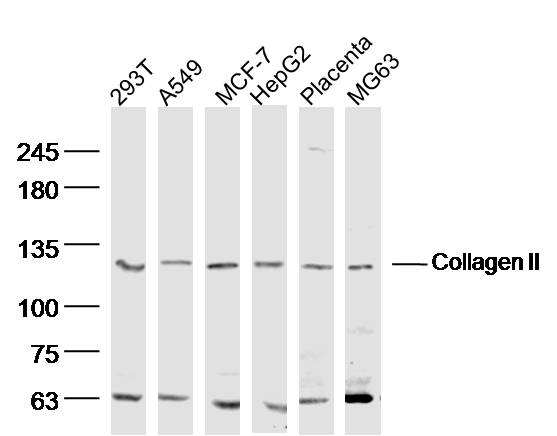 Sample:
293T (human)Cell Lysate at 40 ug
A549 (human)Cell Lysate at 40 ug
MCF-7 (human)Cell Lysate at 40 ug
HepG2 (human)Cell Lysate at 40 ug
Placenta (mouse) Lysate at 40 ug
MG63 (human)Cell Lysate at 40 ug
Primary: Anti- Collagen II (SL0709R) at 1/300 dilution
Secondary: IRDye800CW Goat Anti-Rabbit IgG at 1/20000 dilution
Predicted band size: 117 kD
Observed band size: 120 kD
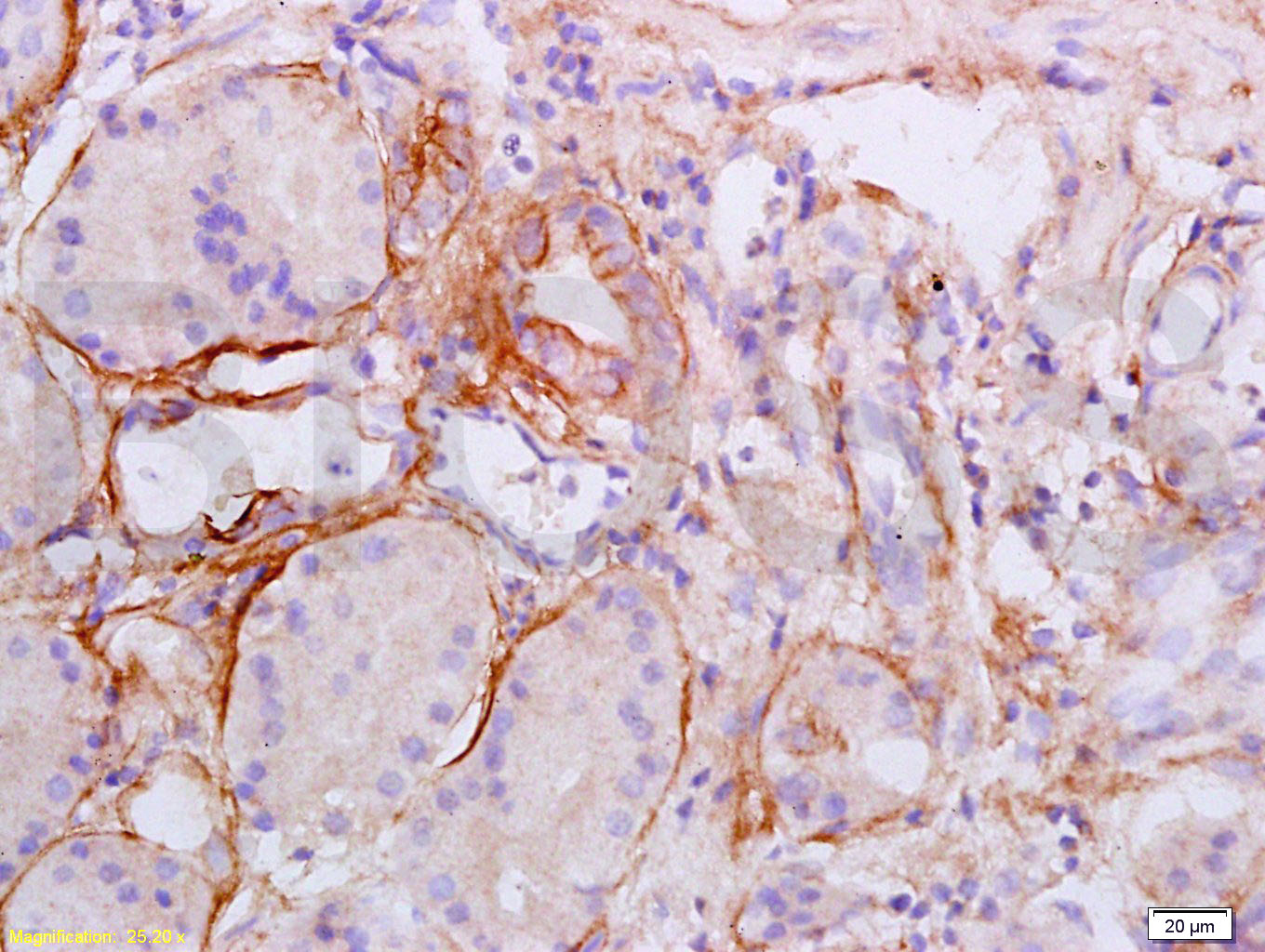 Tissue/cell: human esophageal carcinoma; 4% Paraformaldehyde-fixed and paraffin-embedded;
Antigen retrieval: citrate buffer ( 0.01M, pH 6.0 ), Boiling bathing for 15min; Block endogenous peroxidase by 3% Hydrogen peroxide for 30min; Blocking buffer (normal goat serum,C-0005) at 37℃ for 20 min;
Incubation: Anti-Collagen II Polyclonal Antibody, Unconjugated(SL0709R) 1:200, overnight at 4°C, followed by conjugation to the secondary antibody(SP-0023) and DAB(C-0010) staining
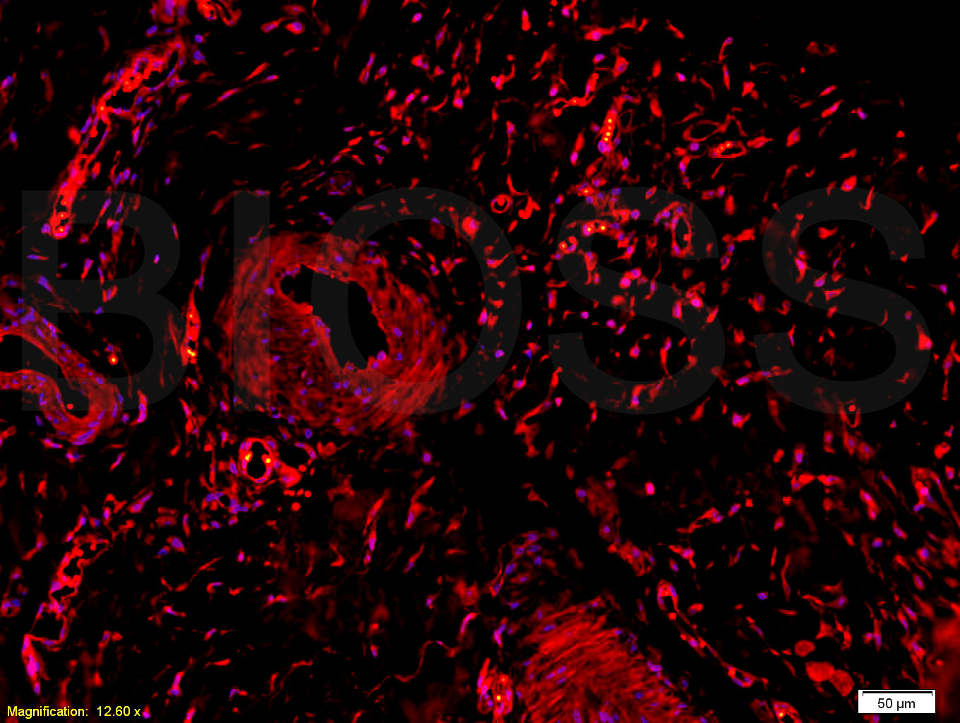 Tissue/cell: rabbit meniscus tissue;4% Paraformaldehyde-fixed and paraffin-embedded;
Antigen retrieval: citrate buffer ( 0.01M, pH 6.0 ), Boiling bathing for 15min; Blocking buffer (normal goat serum,C-0005) at 37℃ for 20 min;
Incubation: Anti-Collagen II Polyclonal Antibody, Unconjugated(SL0709R) 1:200, overnight at 4°C; The secondary antibody was Goat Anti-Rabbit IgG, Cy3 conjugated(SL0295G-Cy3)used at 1:200 dilution for 40 minutes at 37°C. DAPI(5ug/ml,blue,C-0033) was used to stain the cell nuclei
Cartpieces
Totalgoods,subtotals:¥Checkout
Bought notes(bought amounts latest0)
No one bought this product
User Comment(Total0User Comment Num)
- No comment


 +86 571 56623320
 +86 18668110335

