
Rabbit Anti-CD4 antibody
CD4 (L3T4); CD4 antigen (p55); CD4 Antigen ; CD4 molecule; CD4 Receptor; CD4+ Lymphocyte deficiency, included; CD4mut; L3T4; Leu3; Ly-4; Lymphocyte antigen CD4; MGC165891; p55; T Cell Antigen T4 ; T cell antigen T4/LEU3; T cell differentiation antigen L3T
View History [Clear]
Details
Product Name CD4 Chinese Name CD4抗体 Alias CD4 (L3T4); CD4 antigen (p55); CD4 Antigen ; CD4 molecule; CD4 Receptor; CD4+ Lymphocyte deficiency, included; CD4mut; L3T4; Leu3; Ly-4; Lymphocyte antigen CD4; MGC165891; p55; T Cell Antigen T4 ; T cell antigen T4/LEU3; T cell differentiation antigen L3T4; T cell OKT4 deficiency, included; T cell surface antigen T4/Leu 3 ; T cell surface antigen T4/Leu3; T Cell Surface Glycoprotein CD4; W3/25; W3/25 antigen; T-cell surface glycoprotein CD4 isoform 1 precursor; CD4_HUMAN. literatures Research Area Tumour Cell biology immunology Stem cells transcriptional regulatory factor Cell Surface Molecule t-lymphocyte Immunogen Species Rabbit Clonality Polyclonal React Species Human, Mouse, Rat, Pig, (predicted: Dog, Cow, Sheep, Guinea Pig, ) Applications WB=1:500-2000 ELISA=1:5000-10000 IHC-P=1:100-500 IHC-F=1:100-500 Flow-Cyt=1μg/Test IF=1:100-500 (Paraffin sections need antigen repair)
not yet tested in other applications.
optimal dilutions/concentrations should be determined by the end user.Theoretical molecular weight 48kDa Cellular localization The cell membrane Form Liquid Concentration 1mg/ml immunogen KLH conjugated synthetic peptide derived from human CD4: 385-457/457 <Cytoplasmic> Lsotype IgG Purification affinity purified by Protein A Buffer Solution 0.01M TBS(pH7.4) with 1% BSA, 0.03% Proclin300 and 50% Glycerol. Storage Shipped at 4℃. Store at -20 °C for one year. Avoid repeated freeze/thaw cycles. Attention This product as supplied is intended for research use only, not for use in human, therapeutic or diagnostic applications. PubMed PubMed Product Detail This gene encodes a membrane glycoprotein of T lymphocytes that interacts with major histocompatibility complex class II antigenes and is also a receptor for the human immunodeficiency virus. This gene is expressed not only in T lymphocytes, but also in B cells, macrophages, and granulocytes. It is also expressed in specific regions of the brain. The protein functions to initiate or augment the early phase of T-cell activation, and may function as an important mediator of indirect neuronal damage in infectious and immune-mediated diseases of the central nervous system. Multiple alternatively spliced transcript variants encoding different isoforms have been identified in this gene. [provided by RefSeq, Aug 2010].
Function:
Accessory protein for MHC class-II antigen/T-cell receptor interaction. May regulate T-cell activation. Induces the aggregation of lipid rafts.
Subunit:
Associates with LCK. Binds to HIV-1 gp120 and to P4HB/PDI and upon HIV-1 binding to the cell membrane, is part of P4HB/PDI-CD4-CXCR4-gp120 complex. Interacts with HIV-1 Envelope polyprotein gp160 and protein Vpu. Interacts with Human Herpes virus 7 capsid proteins. Interacts with PTK2/FAK1; this interaction requires the presence of HIV-1 gp120.
Subcellular Location:
Cell membrane; Single-pass type I membrane protein. Note=Localizes to lipid rafts. Removed from plasma membrane by HIV-1 Nef protein that increases clathrin-dependent endocytosis of this antigen to target it to lysosomal degradation. Cell surface expression is also down-modulated by HIV-1 Envelope polyprotein gp160 that interacts with, and sequesters CD4 in the endoplasmic reticulum.
Post-translational modifications:
Palmitoylation and association with LCK contribute to the enrichment of CD4 in lipid rafts.
Similarity:
Contains 3 Ig-like C2-type (immunoglobulin-like) domains.
Contains 1 Ig-like V-type (immunoglobulin-like) domain.
SWISS:
P01730
Gene ID:
920
Database links:
Entrez Gene: 920 Human
Entrez Gene: 12504 Mouse
Omim: 186940 Human
SwissProt: P06332 Mouse
SwissProt: P01730 Human
Unigene: 631659 Human
Unigene: 2209 Mouse
此抗体可识别55KDⅠ型单链穿膜glycoprotein。 CD4分子是存在于大多数辅助/诱导T细胞表面的59kDa的glycoprotein。正常淋巴组织中CD4的表达数量多于CD8,此抗体主要用于标记辅助/诱导T细胞,与CD8单抗联合使用对外周血lymphocyte分型。 CD4抗原是HLA-II类分子和人类免疫缺陷病毒(HIV)-爱滋病的受体,在35-50%外周血lymphocyte-辅助和诱导T细胞(Th/Ti)和70-80%人胸腺细胞上表达,在人的单核细胞表面也有低密度的表达。 CD4抗原有膜结合型和可溶性两种形式。Th/Ti可辅助Ig产生和T细胞毒T细胞的作用。Product Picture 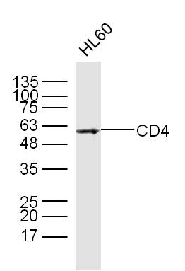 Sample:
HL60 Cell Lysate at 40 ug
Primary: Anti- CD4 (SL0647R) at 1/300 dilution
Secondary: IRDye800CW Goat Anti-Rabbit IgG at 1/20000 dilution
Predicted band size: 48 kD
Observed band size: 55kD
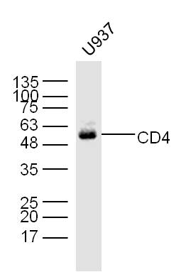 U937 Cell Lysate at 40 ug
Primary: Anti- CD4 (SL0647R) at 1/300 dilution
Secondary: IRDye800CW Goat Anti-Rabbit IgG at 1/20000 dilution
Predicted band size: 48 kD
Observed band size: 55kD
 Sample:
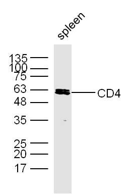
Spleen (Mouse) Lysate at 40 ug
Primary: Anti- CD4 (SL0647R) at 1/300 dilution
Secondary: IRDye800CW Goat Anti-Rabbit IgG at 1/20000 dilution
Predicted band size: 48 kD
Observed band size: 55kD
 Sample:
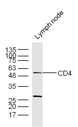
Lymph node(Mouse) Lysate at 40 ug
Primary: Anti- CD4 (SL0647R) at 1/300 dilution
Secondary: IRDye800CW Goat Anti-Rabbit IgG at 1/20000 dilution
Predicted band size: 48 kD
Observed band size: 55kD
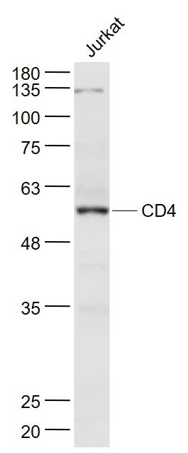 Sample:
Jurkat(Human) Cell Lysate at 30 ug
Primary: Anti- CD4 (SL0647R) at 1/1000 dilution
Secondary: IRDye800CW Goat Anti-Rabbit IgG at 1/20000 dilution
Predicted band size: 48 kD
Observed band size: 55 kD
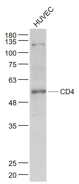 Sample:
HUVEC(Human) Cell Lysate at 30 ug
Primary: Anti- CD4 (SL0647R) at 1/1000 dilution
Secondary: IRDye800CW Goat Anti-Rabbit IgG at 1/20000 dilution
Predicted band size: 48 kD
Observed band size: 55 kD
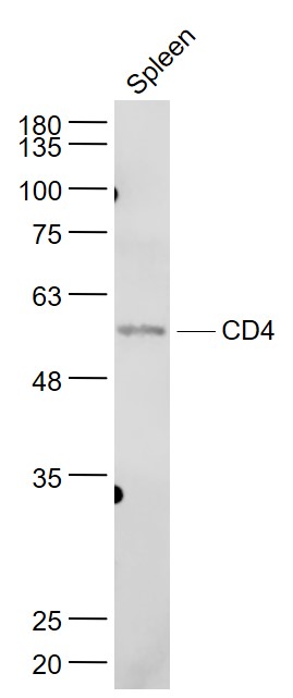 Sample:
Spleen (Mouse) Lysate at 40 ug
Primary: Anti- CD4 (SL0647R) at 1/1000 dilution
Secondary: IRDye800CW Goat Anti-Rabbit IgG at 1/20000 dilution
Predicted band size: 48 kD
Observed band size: 55 kD
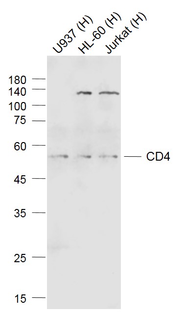 Sample:
Lane 1: U937 (Human) Cell Lysate at 30 ug
Lane 2: HL-60 (Human) Cell Lysate at 30 ug
Lane 3: Jurkat (Human) Cell Lysate at 30 ug
Primary: Anti-CD4 (SL0647R) at 1/1000 dilution
Secondary: IRDye800CW Goat Anti-Rabbit IgG at 1/20000 dilution
Predicted band size: 55 kD
Observed band size: 55 kD
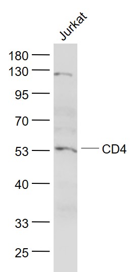 Sample:
Jurkat(Human) Cell Lysate at 30 ug
Primary: Anti- CD4 (SL0647R) at 1/1000 dilution
Secondary: IRDye800CW Goat Anti-Rabbit IgG at 1/20000 dilution
Predicted band size: 48 kD
Observed band size: 53 kD
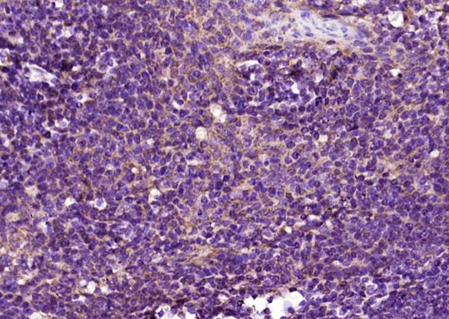 Paraformaldehyde-fixed, paraffin embedded (human tonsil); Antigen retrieval by boiling in sodium citrate buffer (pH6.0) for 15min; Block endogenous peroxidase by 3% hydrogen peroxide for 20 minutes; Blocking buffer (normal goat serum) at 37°C for 30min; Antibody incubation with (CD4) Polyclonal Antibody, Unconjugated (SL0647R) at 1:4000 overnight at 4°C, followed by operating according to SP Kit(Rabbit) (sp-0023) instructionsand DAB staining.
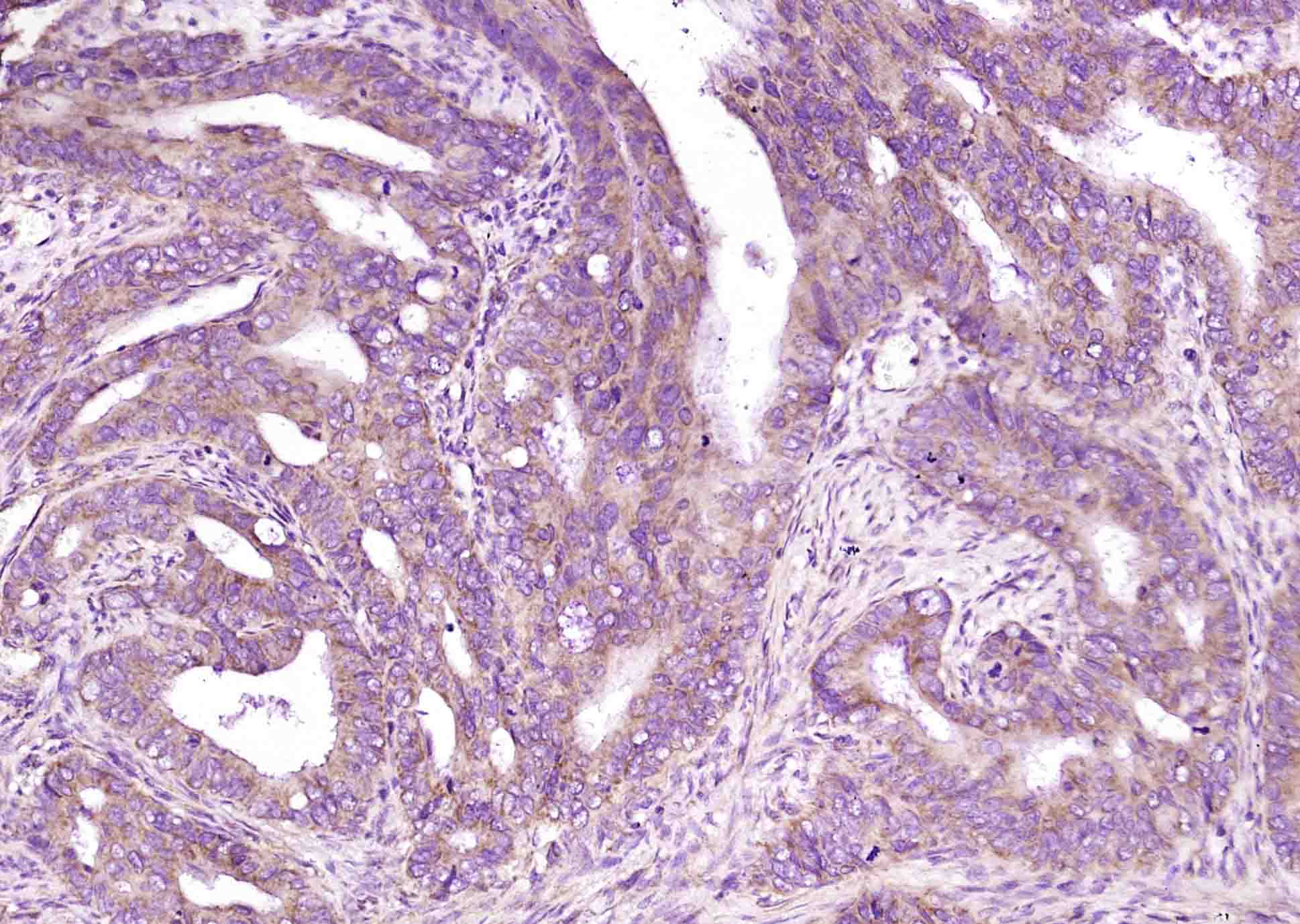 Paraformaldehyde-fixed, paraffin embedded (human colon carcinoma); Antigen retrieval by boiling in sodium citrate buffer (pH6.0) for 15min; Block endogenous peroxidase by 3% hydrogen peroxide for 20 minutes; Blocking buffer (normal goat serum) at 37°C for 30min; Antibody incubation with (CD4) Polyclonal Antibody, Unconjugated (SL0647R) at 1:4000 overnight at 4°C, followed by operating according to SP Kit(Rabbit) (sp-0023) instructionsand DAB staining.
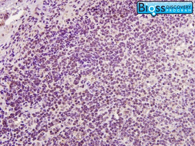 Image was kindly submitted by Dr.David M Burmeister from US Army Institute of Surgical Research. Pig lymph nodes stained with Rabbit Anti-CD4 Polyclonal Antibody(SLSL0647R)at 1:300 for one hour at room temperature.
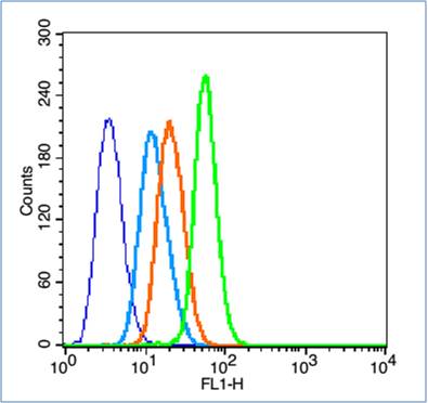 Blank control (blue line): HL60 (fixed with 2% paraformaldehyde (10 min) and then permeabilized with 0.1% PBS-Tween for 20 min at room temperature).
Primary Antibody (green line): Rabbit Anti-CD4 antibody (SL0647R),Dilution: 1μg /10^6 cells;
Isotype Control Antibody (orange line): Rabbit IgG .
Secondary Antibody (white blue line): Goat anti-rabbit IgG-FITC,dilution: 1μg /test.
Cartpieces
Totalgoods,subtotals:¥Checkout
Bought notes(bought amounts latest0)
No one bought this product
User Comment(Total0User Comment Num)
- No comment


 +86 571 56623320
 +86 18668110335

