
Rabbit Anti-CD34 antibody
CD34 antigen; CD34 molecule; Cluster designation 34; Hematopoietic progenitor cell antigen CD34; HPCA1; CD34_HUMAN.
View History [Clear]
Details
Product Name CD34 Chinese Name CD34抗体 Alias CD34 antigen; CD34 molecule; Cluster designation 34; Hematopoietic progenitor cell antigen CD34; HPCA1; CD34_HUMAN. literatures Research Area Tumour Cardiovascular immunology Developmental biology Neurobiology Stem cells Cell Surface Molecule glycoprotein Cell type markers vascular endothelial cell endothelial cells Immunogen Species Rabbit Clonality Polyclonal React Species Human, Mouse, (predicted: Rat, Dog, Pig, Cow, Rabbit, ) Applications WB=1:500-2000 ELISA=1:5000-10000 IHC-P=1:100-500 IHC-F=1:100-500 Flow-Cyt=1μg/Test IF=1:200-800 (Paraffin sections need antigen repair)
not yet tested in other applications.
optimal dilutions/concentrations should be determined by the end user.Theoretical molecular weight 39kDa Cellular localization The cell membrane Form Liquid Concentration 1mg/ml immunogen KLH conjugated synthetic peptide derived from human CD34: 301-385/385 <Extracellular> Lsotype IgG Purification affinity purified by Protein A Buffer Solution 0.01M TBS(pH7.4) with 1% BSA, 0.03% Proclin300 and 50% Glycerol. Storage Shipped at 4℃. Store at -20 °C for one year. Avoid repeated freeze/thaw cycles. Attention This product as supplied is intended for research use only, not for use in human, therapeutic or diagnostic applications. PubMed PubMed Product Detail The highly glycosylated 75-120 kD antigen CD34 is possibly an adhesion molecule with a putative role in early hematopoiesis by mediating the attachment of stem cells to the bone marrow extracellular matrix or directly to stromal cells. It could act as a scaffold for the attachment of lineage specific glycans, allowing stem cells to bind to lectins expressed by stromal cells or other marrow components. CD34 is thought to have a role in presenting carbohydrate ligands to selectins. The intracellular chain of the CD34 antigen is a site of phosphorylation by activated protein kinase C, suggesting a putative role in signal transduction. Two isoforms of CD34 have been reported to be generated by alternative splicing. CD34 is highly expressed on hematopoietic progenitors, as well as on endothelial cells, brain, and testis. Staining for CD34 has been used to measure angiogenesis, which reportedly predicts tumor recurrence.
Function:
Possible adhesion molecule with a role in early hematopoiesis by mediating the attachment of stem cells to the bone marrow extracellular matrix or directly to stromal cells. Could act as a scaffold for the attachment of lineage specific glycans, allowing stem cells to bind to lectins expressed by stromal cells or other marrow components. Presents carbohydrate ligands to selectins.
Subcellular Location:
Membrane; Single-pass type I membrane protein.
Tissue Specificity:
Selectively expressed on hematopoietic progenitor cells and the small vessel endothelium of a variety of tissues.
Post-translational modifications:
Highly glycosylated.
Phosphorylated on serine residues by PKC.
Similarity:
Belongs to the CD34 family.
SWISS:
P28906
Gene ID:
947
Database links:Entrez Gene: 947 Human
Omim: 142230 Human
SwissProt: P28906 Human
Unigene: 374990 Human
造血Stem cellsMaker
内皮Maker
Tumour生物Maker
细胞表面的唾液粘蛋白。
间充质Stem cells(mesenchymal stem cells,MSC)也是Stem cells家族的重要成员,来源于发育早期的中胚层和外胚层。MSC最初在骨髓中发现,因其具有多向分化潜能、造血支持和促进Stem cells植入、免疫调控和自我复制等特点。如间充质Stem cells在体内或体外特定的诱导条件下,可分化为血管内皮、脂肪、骨、软骨、肌肉、肌腱、韧带、神经、肝、心肌、等多种组织细胞。Product Picture 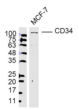 Sample:
MCF-7(Human) Cell Lysate at 40 ug
Primary: Anti-CD34 (SL0646R) at 1/300 dilution
Secondary: IRDye800CW Goat Anti-Rabbit IgG at 1/20000 dilution
Predicted band size: 39 kD
Observed band size: 115 kD
 Sample:
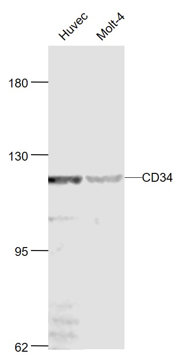
Huvec(Human) Cell Lysate at 30 ug
Molt-4(Human) Cell Lysate at 30 ug
Primary: Anti-CD34 (SL0646R) at 1/1000 dilution
Secondary: IRDye800CW Goat Anti-Rabbit IgG at 1/20000 dilution
Predicted band size: 120 kD
Observed band size: 120 kD
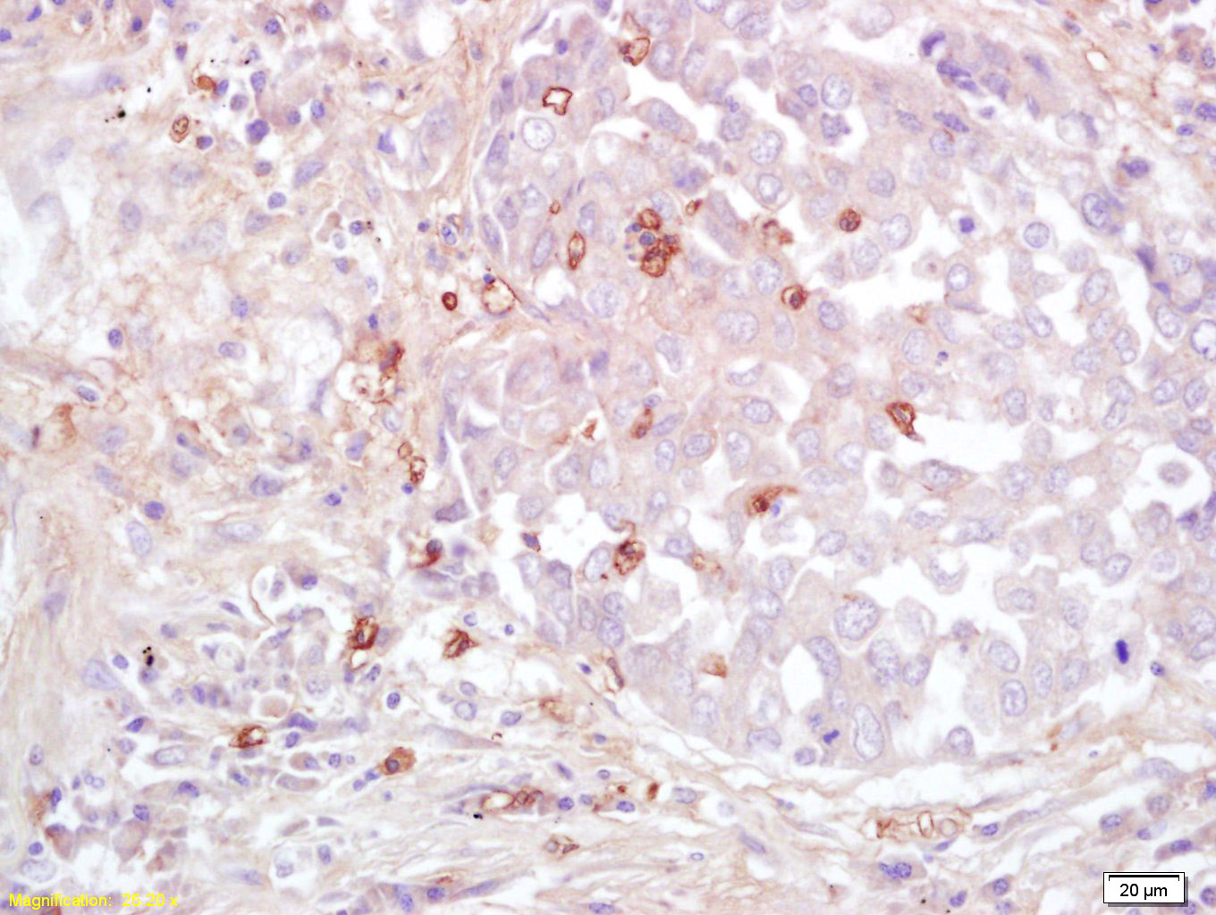 Tissue/cell: human lung carcinoma; 4% Paraformaldehyde-fixed and paraffin-embedded;
Antigen retrieval: citrate buffer ( 0.01M, pH 6.0 ), Boiling bathing for 15min; Block endogenous peroxidase by 3% Hydrogen peroxide for 30min; Blocking buffer (normal goat serum,C-0005) at 37℃ for 20 min;
Incubation: Anti-CD34 Polyclonal Antibody, Unconjugated(SL0646R) 1:200, overnight at 4°C, followed by conjugation to the secondary antibody(SP-0023) and DAB(C-0010) staining
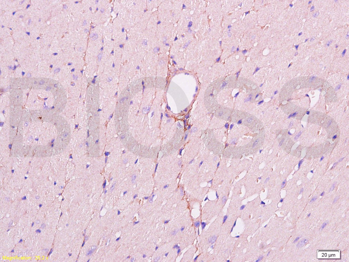 Tissue/cell: mouse heart tissue; 4% Paraformaldehyde-fixed and paraffin-embedded;
Antigen retrieval: citrate buffer ( 0.01M, pH 6.0 ), Boiling bathing for 15min; Block endogenous peroxidase by 3% Hydrogen peroxide for 30min; Blocking buffer (normal goat serum,C-0005) at 37℃ for 20 min;
Incubation: Anti-CD34 Polyclonal Antibody, Unconjugated(SL0646R) 1:200, overnight at 4°C, followed by conjugation to the secondary antibody(SP-0023) and DAB(C-0010) staining
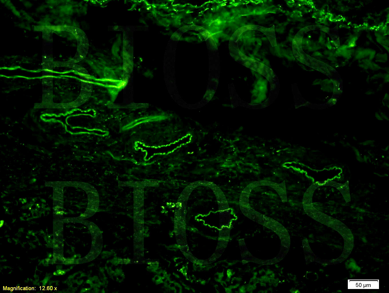 Tissue/cell: human gastric adenocarcinoma;4% Paraformaldehyde-fixed and paraffin-embedded;
Antigen retrieval: citrate buffer ( 0.01M, pH 6.0 ), Boiling bathing for 15min; Blocking buffer (normal goat serum,C-0005) at 37℃ for 20 min;
Incubation: Anti-CD34 Polyclonal Antibody, Unconjugated(SL0646R) 1:200, overnight at 4°C; The secondary antibody was Goat Anti-Rabbit IgG, AF488 conjugated(SL0295G-AF488)used at 1:200 dilution for 40 minutes at 37°C.
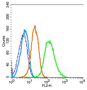 Blank control: Raji(blue).
Primary Antibody:Rabbit Anti- CD34 antibody(SL0646R), Dilution: 1μg in 100 μL 1X PBS containing 0.5% BSA;
Isotype Control Antibody: Rabbit IgG(orange) ,used under the same conditions );
Secondary Antibody: Goat anti-rabbit IgG-PE(white blue), Dilution: 1:200 in 1 X PBS containing 0.5% BSA.
Protocol
The cells were fixed with 2% paraformaldehyde (10 min). Antibody (SL0646R, 1μg /1x10^6 cells) were incubated for 30 min on the ice, followed by 1 X PBS containing 0.5% BSA + 1 0% goat serum (15 min) to block non-specific protein-protein interactions. Then the Goat Anti-rabbit IgG/PE antibody was added into the blocking buffer mentioned above to react with the primary antibody of SL0646R at 1/200 dilution for 30 min on ice. Acquisition of 20,000 events was performed.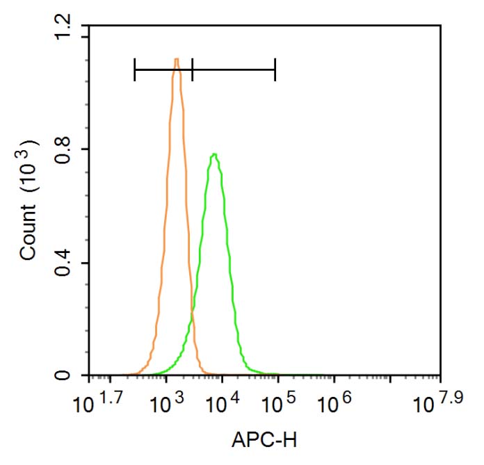 Blank control: HUVEC.
Primary Antibody (green line): Rabbit CD34 antibody (SL0646R)
Dilution: 1μg /10^6 cells;
Isotype Control Antibody (orange line): Rabbit IgG .
Secondary Antibody: Goat anti-rabbit IgG-AF647
Dilution: 1μg /test.
Protocol
The cells were then incubated in 5%BSA to block non-specific protein-protein interactions for 30 min at room temperature .Cells stained with Primary Antibody for 30 min at room temperature. The secondary antibody used for 40 min at room temperature. Acquisition of 20,000 events was performed.
Cartpieces
Totalgoods,subtotals:¥Checkout
Bought notes(bought amounts latest0)
No one bought this product
User Comment(Total0User Comment Num)
- No comment


 +86 571 56623320
 +86 18668110335

