
Rabbit Anti-VCAM1 antibody
VCAM1; CD 106; CD106; CD106 Antigen; DKFZp779G2333; INCAM 100; L1CAM; MGC99561; Vascular Cell Adhesion Molecule 1; Vascular cell adhesion protein 1; VCAM 1; VCAM-1; INCAM-100; V-CAM 1; VCAM1_HUMAN.
View History [Clear]
Details
Product Name VCAM1 Chinese Name vascular endothelial cell粘附分子(CD106)抗体 Alias VCAM1; CD 106; CD106; CD106 Antigen; DKFZp779G2333; INCAM 100; L1CAM; MGC99561; Vascular Cell Adhesion Molecule 1; Vascular cell adhesion protein 1; VCAM 1; VCAM-1; INCAM-100; V-CAM 1; VCAM1_HUMAN. literatures Research Area Tumour Cardiovascular Cell biology Cell adhesion molecule endothelial cells Immunogen Species Rabbit Clonality Polyclonal React Species Human, Mouse, (predicted: Rat, ) Applications WB=1:500-2000 ELISA=1:5000-10000 IHC-P=1:100-500 IHC-F=1:100-500 Flow-Cyt=1µg/Test IF=1:100-500 (Paraffin sections need antigen repair)
not yet tested in other applications.
optimal dilutions/concentrations should be determined by the end user.Theoretical molecular weight 81kDa Cellular localization The cell membrane Form Liquid Concentration 1mg/ml immunogen KLH conjugated synthetic peptide derived from mouse VCAM-1: 640-739/739 <Extracellular> Lsotype IgG Purification affinity purified by Protein A Buffer Solution 0.01M TBS(pH7.4) with 1% BSA, 0.03% Proclin300 and 50% Glycerol. Storage Shipped at 4℃. Store at -20 °C for one year. Avoid repeated freeze/thaw cycles. Attention This product as supplied is intended for research use only, not for use in human, therapeutic or diagnostic applications. PubMed PubMed Product Detail VCAM1 is important in cell-cell recognition. Appears to function in leukocyte-endothelial cell adhesion. Interacts with the integrins alpha4 beta1 (beta 1 integrin VLA4) and alpha4 beta7 on leukocytes, and mediates both adhesion and signal transduction. The VCAM1/VLA4 interaction may play a pathophysiologic role both in immune responses and in leukocyte emigration to sites of inflammation. VCAM1 is also expressed by several non endothelial cell types including some macrophages, follicular dendritic cells and bone marrow, stromal cells.
Function:
Important in cell-cell recognition. Appears to function in leukocyte-endothelial cell adhesion. Interacts with the beta-1 integrin VLA4 on leukocytes, and mediates both adhesion and signal transduction. The VCAM1/VLA4 interaction may play a pathophysiologic role both in immune responses and in leukocyte emigration to sites of inflammation.
Subunit:
Binds to ECMV-D capsid proteins and acts as a receptor for this virus.
Subcellular Location:
Isoform 1: Cell membrane; Single-pass type I membrane protein. Isoform 2: Cell membrane; Lipid-anchor, GPI-anchor.
Tissue Specificity:
Expressed on inflamed vascular endothelium, as well as on macrophage-like and dendritic cell types in both normal and inflamed tissue. Expressed in the bone marrow.
Similarity:
Contains 7 Ig-like C2-type (immunoglobulin-like) domains.
SWISS:
P29533
Gene ID:
22329
Database links:Entrez Gene: 7412 Human
Entrez Gene: 22329 Mouse
GenBank: NP_001069.1 Human
GenBank: NP_001186763.1 Human
Omim: 192225 Human
SwissProt: P19320 Human
SwissProt: P29533 Mouse
VCAM 1(CD106)是一种膜相关蛋白,分布于血管内皮及其周围的lymphocyte、单核细胞、嗜酸性粒细胞和中性粒细胞。Anti-VCAM-1主要用于各种组织中血管内皮的检测。Product Picture 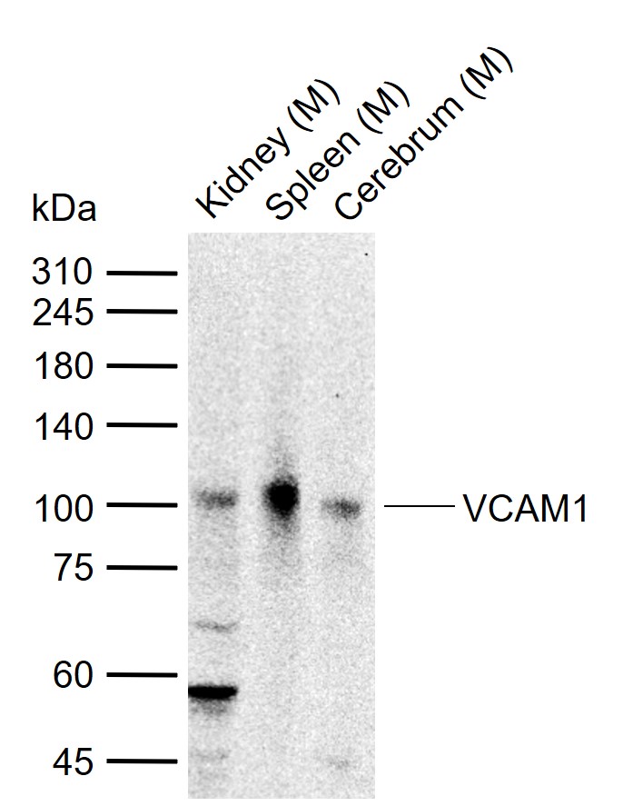 Sample:
Lane 1: Mouse Kidney tissue lysates
Lane 2: Mouse Spleen tissue lysates
Lane 3: Mouse Cerebrum tissue lysates
Primary: Anti-VCAM1 (SL0396R) at 1/500 dilution
Secondary: IRDye800CW Goat Anti-Rabbit IgG at 1/20000 dilution
Predicted band size: 81 kDa
Observed band size: 100 kDa
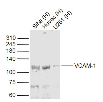 Sample:
Lane 1: Siha (Human) Cell Lysate at 30 ug
Lane 2: Huvec (Human) Cell Lysate at 30 ug
Lane 3: U251 (Human) Cell Lysate at 30 ug
Primary: Anti-VCAM-1 (SL0396R) at 1/1000 dilution
Secondary: IRDye800CW Goat Anti-Rabbit IgG at 1/20000 dilution
Predicted band size: 110 kD
Observed band size: 110 kD
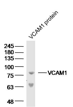 Sample: VCAM1 protein (Human) at 100 ng
Primary: Anti-VCAM-1 (SL0396R) at 1/300 dilution
Secondary: IRDye800CW Goat Anti-Rabbit IgG at 1/20000 dilution
Predicted band size: 81 kD
Observed band size: 81 kD
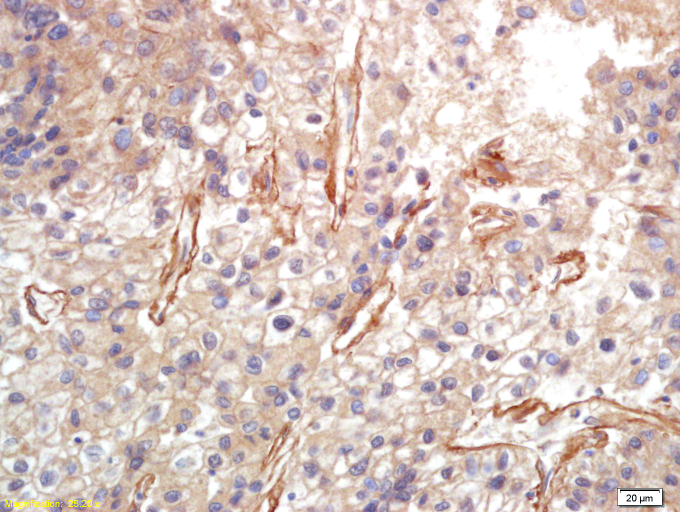 Tissue/cell: human lung carcinoma; 4% Paraformaldehyde-fixed and paraffin-embedded;
Antigen retrieval: citrate buffer ( 0.01M, pH 6.0 ), Boiling bathing for 15min; Block endogenous peroxidase by 3% Hydrogen peroxide for 30min; Blocking buffer (normal goat serum,C-0005) at 37℃ for 20 min;
Incubation: Anti-VCAM-1 Polyclonal Antibody, Unconjugated(SL0396R) 1:200, overnight at 4°C, followed by conjugation to the secondary antibody(SP-0023) and DAB(C-0010) staining
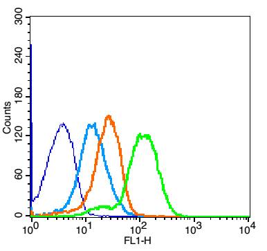 Blank control: Mouse Spleen(blue).
Primary Antibody:Rabbit Anti-VCAM-1 antibody (SL0396R,Green); Dilution: 1μg in 100 μL 1X PBS containing 0.5% BSA;
Isotype Control Antibody: Rabbit IgG(orange) ,used under the same conditions;
Secondary Antibody: Goat anti-rabbit IgG-FITC(white blue), Dilution: 1:200 in 1 X PBS containing 0.5% BSA.
Protocol
The cells were fixed with 2% paraformaldehyde for 10 min at 37℃. Primary antibody (SL0396R, 1μg /8x10^5 cells) were incubated for 30 min at room temperature, followed by 1 X PBS containing 0.5% BSA + 1 0% goat serum (1 hour) to block non-specific protein-protein interactions. Then the Goat Anti-rabbit IgG/FITC antibody was added into the blocking buffer mentioned above to react with the primary antibody at 1/200 dilution for 40 min at room temperature. Acquisition of 20,000 events was performed.
Cartpieces
Totalgoods,subtotals:¥Checkout
Partial purchase records(bought amounts latest0)
No one bought this product
User Comment(Total0User Comment Num)
- No comment


 +86 571 56623320


