
Rabbit Anti-CD133 antibody
AC133; Antigen AC133; Hematopoietic stem cell antigen; hProminin; PROM1; Prominin I; Prominin like protein 1 precursor; Prominin mouse like 1; prominin1; PROML1; CORD12; MCDR2; MSTP061; PROML1; RP41; STGD4; PROM1_HUMAN; CD133 antigen.
View History [Clear]
Details
Product Name CD133 Chinese Name 造血Stem cells抗原CD133抗体 Alias AC133; Antigen AC133; Hematopoietic stem cell antigen; hProminin; PROM1; Prominin I; Prominin like protein 1 precursor; Prominin mouse like 1; prominin1; PROML1; CORD12; MCDR2; MSTP061; PROML1; RP41; STGD4; PROM1_HUMAN; CD133 antigen. literatures Research Area Tumour Cell biology immunology Stem cells Cell type markers Immunogen Species Rabbit Clonality Polyclonal React Species Mouse, (predicted: Human, ) Applications WB=1:500-2000 ELISA=1:5000-10000 Flow-Cyt=1μg /test
not yet tested in other applications.
optimal dilutions/concentrations should be determined by the end user.Theoretical molecular weight 95kDa Detection molecular weight 120 kDa Cellular localization The cell membrane Form Liquid Concentration 1mg/ml immunogen KLH conjugated synthetic peptide derived from human CD133: 751-848/865 <Cytoplasmic> Lsotype IgG Purification affinity purified by Protein A Buffer Solution 0.01M TBS(pH7.4) with 1% BSA, 0.03% Proclin300 and 50% Glycerol. Storage Shipped at 4℃. Store at -20 °C for one year. Avoid repeated freeze/thaw cycles. Attention This product as supplied is intended for research use only, not for use in human, therapeutic or diagnostic applications. PubMed PubMed Product Detail This gene encodes a pentaspan transmembrane glycoprotein. The protein localizes to membrane protrusions and is often expressed on adult stem cells, where it is thought to function in maintaining stem cell properties by suppressing differentiation. Mutations in this gene have been shown to result in retinitis pigmentosa and Stargardt disease. Expression of this gene is also associated with several types of cancer. This gene is expressed from at least five alternative promoters that are expressed in a tissue-dependent manner. Multiple transcript variants encoding different isoforms have been found for this gene. [provided by RefSeq, Mar 2009]
Function:
Binds cholesterol in cholesterol-containing plasma membrane microdomains. Proposed to play a role in apical plasma membrane organization of epithelial cells. During early retinal development acts as a key regulator of disk morphogenesis. Involved in regulation of MAPK and Akt signaling pathways. In neuroblastoma cells suppresses cell differentiation such as neurite outgrowth in a RET-dependent manner.
Subunit:
Interacts with CDHR1 and with actin filaments.
Subcellular Location:
Cell projection, cilium, photoreceptor outer segment. Isoform 1: Apical cell membrane; Multi-pass membrane protein. Cell projection, microvillus membrane; Multi-pass membrane protein. Note=Found in extracellular membrane particles in various body fluids such as cerebrospinal fluid, saliva, seminal fluid and urine.
Tissue Specificity:
Isoform 1 is selectively expressed on CD34 hematopoietic stem and progenitor cells in adult and fetal bone marrow, fetal liver, cord blood and adult peripheral blood. Isoform 1 is not detected on other blood cells. Isoform 1 is also expressed in a number of non-lymphoid tissues including retina, pancreas, placenta, kidney, liver, lung, brain and heart. Found in saliva within small membrane particles. Isoform 2 is predominantly expressed in fetal liver, skeletal muscle, kidney, and heart as well as adult pancreas, kidney, liver, lung, and placenta. Isoform 2 is highly expressed in fetal liver, low in bone marrow, and barely detectable in peripheral blood. Isoform 2 is expressed on hematopoietic stem cells and in epidermal basal cells (at protein level). Expressed in adult retina by rod and cone photoreceptor cells (at protein level).
Post-translational modifications:
Isoform 1 and isoform 2 are glycosylated.
DISEASE:
Defects in PROM1 are the cause of retinitis pigmentosa type 41 (RP41) [MIM:612095]; also known as retinal degeneration autosomal recessive prominin-related. RP is a retinal dystrophy belonging to the group of pigmentary retinopathies. RP is characterized by retinal pigment deposits visible on fundus examination and primary loss of rod photoreceptor cells followed by secondary loss of cone photoreceptors. Patients typically have night vision blindness and loss of midperipheral visual field. As their condition progresses, they lose their far peripheral visual field and eventually central vision as well.
Defects in PROM1 are the cause of cone-rod dystrophy type 12 (CORD12) [MIM:612657]. CORD12 is an inherited retinal dystrophy characterized by retinal pigment deposits visible on fundus examination, predominantly in the macular region, and initial loss of cone photoreceptors followed by rod degeneration. This leads to decreased visual acuity and sensitivity in the central visual field, followed by loss of peripheral vision. Severe loss of vision occurs earlier than in retinitis pigmentosa.
Defects in PROM1 are the cause of Stargardt disease type 4 (STGD4) [MIM:603786]. Stargardt disease is the most common hereditary macular degeneration. It is characterized by decreased central vision, atrophy of the macula and underlying retinal pigment epithelium, and frequent presence of prominent flecks in the posterior pole of the retina.
Defects in PROM1 are the cause of retinal macular dystrophy type 2 (MCDR2) [MIM:608051]. MCDR2 is a bull's-eye macular dystrophy characterized by bilateral annular atrophy of retinal pigment epithelium at the macula.
Similarity:
Belongs to the prominin family.
SWISS:
O43490
Gene ID:
8842
Database links:Entrez Gene: 8842 Human
Entrez Gene: 19126 Mouse
Omim: 604365 Human
SwissProt: O43490 Human
SwissProt: O54990 Mouse
Unigene: 614734 Human
Unigene: 6250 Mouse
Unigene: 144589 Rat
Stem cellsMaker
一般认为,VEGFR2(血管内皮生长因子受体2)是HSCs(造血Stem cells)的特异性的表面标志。近来经研究发现CD133分子是HSCs(造血Stem cells)特异性标志。CD133即AC133,是一个新发现的HSCs(造血Stem cells)表面标志,在HSCs(造血Stem cells)分化成熟过程中,CD133的含量迅速降低。EPCs(血管内皮前体细胞)区别于成熟endothelial cells的主要标志是CD133。
经研究发现endothelial cells不能结合CD133的抗体。证实分化成熟的endothelial cells不具有CD133。这些说明CD133可以作为EPCs(血管内皮前体细胞)区别于成熟endothelial cells的一个表面标志.Product Picture 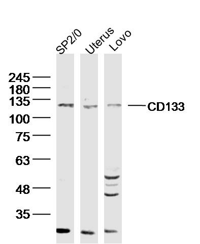 Sample:
SP2/0 (mouse)cell Lysate at 40 ug
Uterus (mouse) Lysate at 40 ug
Lovo (human)cell Lysate at 40 ug
Primary: Anti- CD133 (SL0395R)at 1/300 dilution
Secondary: IRDye800CW Goat Anti-Rabbit IgG at 1/20000 dilution
Predicted band size: 95kD
Observed band size: 115 kD
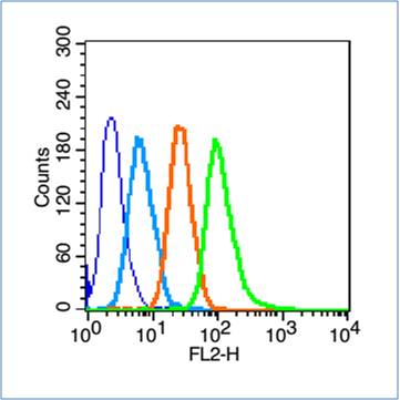 Blank control (blue line): Hep G2(blue).
Primary Antibody (green line): Rabbit Anti-CD133 antibody (SL0395R)
Dilution: 1μg /10^6 cells;
Isotype Control Antibody (orange line): Rabbit IgG .
Secondary Antibody (white blue line): Goat anti-rabbit IgG-PE
Dilution: 1μg /test.
Protocol
The cells were fixed with 70% ethanol Overnight at 4℃. Cells stained with Primary Antibody for 30 min at room temperature. The cells were then incubated in 1 X PBS/2%BSA/10% goat serum to block non-specific protein-protein interactions followed by the antibody for 15 min at room temperature. The secondary antibody used for 40 min at room temperature. Acquisition of 20,000 events was performed.
Cartpieces
Totalgoods,subtotals:¥Checkout
Partial purchase records(bought amounts latest0)
No one bought this product
User Comment(Total0User Comment Num)
- No comment


 +86 571 56623320


