
Rabbit Anti-Caspase-6 subunit p18 antibody
Caspase-6; Caspase-6 p18; cleaved Caspase-6 p18; Apoptosis Related Cysteine Protease; Apoptotic cysteine protease MCH2; Apoptotic protease MCH 2; Apoptotic protease MCH2; Casp 6; Casp6; Caspase 6 apoptosis related cysteine protease; Caspase 6 precursor; C
View History [Clear]
Details
Product Name Caspase-6 subunit p18 Chinese Name 半胱胺酸蛋白酶蛋白-6抗体 Alias Caspase-6; Caspase-6 p18; cleaved Caspase-6 p18; Apoptosis Related Cysteine Protease; Apoptotic cysteine protease MCH2; Apoptotic protease MCH 2; Apoptotic protease MCH2; Casp 6; Casp6; Caspase 6 apoptosis related cysteine protease; Caspase 6 precursor; Caspase6; Human cysteine protease Mch2 isoform alpha; Mch 2; Mch2; CASP6_HUMAN; caspase-6 isoform alpha precursor. Research Area Cell biology Neurobiology Signal transduction Apoptosis Immunogen Species Rabbit Clonality Polyclonal React Species Human, Mouse, (predicted: Rat, Dog, Rabbit, ) Applications WB=1:500-2000 ELISA=1:5000-10000 IHC-P=1:100-500 IHC-F=1:100-500 IF=1:100-500 (Paraffin sections need antigen repair)
not yet tested in other applications.
optimal dilutions/concentrations should be determined by the end user.Theoretical molecular weight 18/33kDa Cellular localization cytoplasmic Form Liquid Concentration 1mg/ml immunogen KLH conjugated synthetic peptide derived from human Caspase-6 subunit p18: 27-130/293 Lsotype IgG Purification affinity purified by Protein A Buffer Solution 0.01M TBS(pH7.4) with 1% BSA, 0.03% Proclin300 and 50% Glycerol. Storage Shipped at 4℃. Store at -20 °C for one year. Avoid repeated freeze/thaw cycles. Attention This product as supplied is intended for research use only, not for use in human, therapeutic or diagnostic applications. PubMed PubMed Product Detail This gene encodes a protein which is a member of the cysteine-aspartic acid protease (caspase) family. Sequential activation of caspases plays a central role in the execution-phase of cell apoptosis. Caspases exist as inactive proenzymes which undergo proteolytic processing at conserved aspartic residues to produce two subunits, large and small, that dimerize to form the active enzyme. This protein is processed by caspases 7, 8 and 10, and is thought to function as a downstream enzyme in the caspase activation cascade. Alternative splicing of this gene results in two transcript variants that encode different isoforms. [provided by RefSeq, Jul 2008]
Function:
Involved in the activation cascade of caspases responsible for apoptosis execution. Cleaves poly(ADP-ribose) polymerase in vitro, as well as lamins. Overexpression promotes programmed cell death.
Subunit:
Heterotetramer that consists of two anti-parallel arranged heterodimers, each one formed by a 18 kDa (p18) and a 11 kDa (p11) subunit. Interacts with BIRC6/bruce.
Subcellular Location:
Cytoplasm.
Post-translational modifications:
Cleavages by caspase-3, caspase-8 or -10 generate the two active subunits.
Similarity:
Belongs to the peptidase C14A family.
SWISS:
P55212
Gene ID:
839
Database links:Entrez Gene: 839 Human
Entrez Gene: 12368 Mouse
Omim: 601532 Human
SwissProt: P55212 Human
SwissProt: O08738 Mouse
Unigene: 654616 Human
Unigene: 281379 Mouse
Unigene: 88160 Rat
Caspase-6 与Caspase-3有38%的同源性,属胱氨酸-天冬氨酸蛋白酶家族。在凋亡执行阶段起中心作用,可被Caspase-7、-8、-10剪切。它是唯一一个可以剪切核纤层蛋白的Caspase。Product Picture 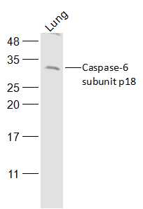 Sample:
Lung (Mouse) Lysate at 40 ug
Primary: Anti-Caspase-6 subunit p18 (SL0084R) at 1/500 dilution
Secondary: IRDye800CW Goat Anti-Rabbit IgG at 1/20000 dilution
Predicted band size: 18/33 kD
Observed band size: 33 kD
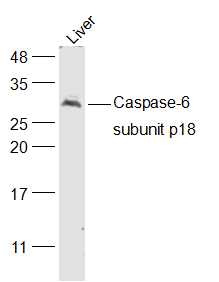 Sample:
Liver (Mouse) Lysate at 40 ug
Primary: Anti-Caspase-6 subunit p18 (SL0084R) at 1/500 dilution
Secondary: IRDye800CW Goat Anti-Rabbit IgG at 1/20000 dilution
Predicted band size: 18/33 kD
Observed band size: 33 kD
 Tissue/cell: Human kidney tissue; 4% Paraformaldehyde-fixed and paraffin-embedded;
Antigen retrieval: citrate buffer ( 0.01M, pH 6.0 ), Boiling bathing for 15min; Block endogenous peroxidase by 3% Hydrogen peroxide for 30min; Blocking buffer (normal goat serum,C-0005) at 37℃ for 20 min;
Incubation: Anti- Caspase-6 Polyclonal Antibody, Unconjugated(SL0084R) 1:200, overnight at 4°C, followed by conjugation to the secondary antibody(SP-0023) and DAB(C-0010) staining
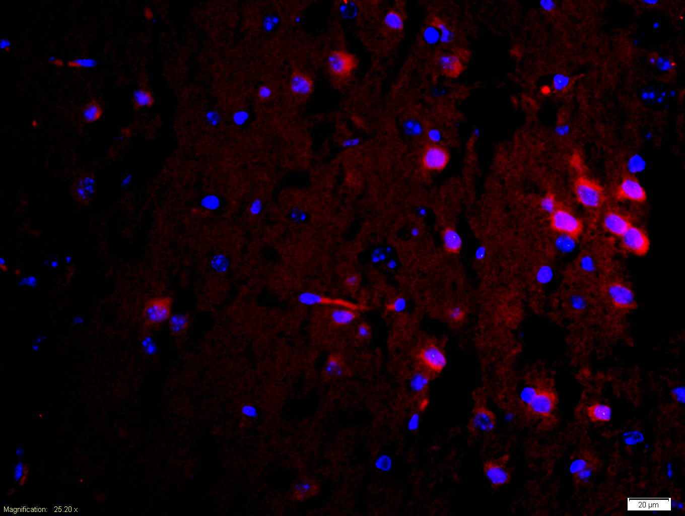 Tissue/cell: mouse brain tissue;4% Paraformaldehyde-fixed and paraffin-embedded;
Antigen retrieval: citrate buffer ( 0.01M, pH 6.0 ), Boiling bathing for 15min; Blocking buffer (normal goat serum,C-0005) at 37℃ for 20 min;
Incubation: Anti-Caspase-6 Polyclonal Antibody, Unconjugated(SL0084R) 1:200, overnight at 4°C; The secondary antibody was Goat Anti-Rabbit IgG, Cy3 conjugated(SL0295G-Cy3)used at 1:200 dilution for 40 minutes at 37°C. DAPI(5ug/ml,blue,C-0033) was used to stain the cell nuclei
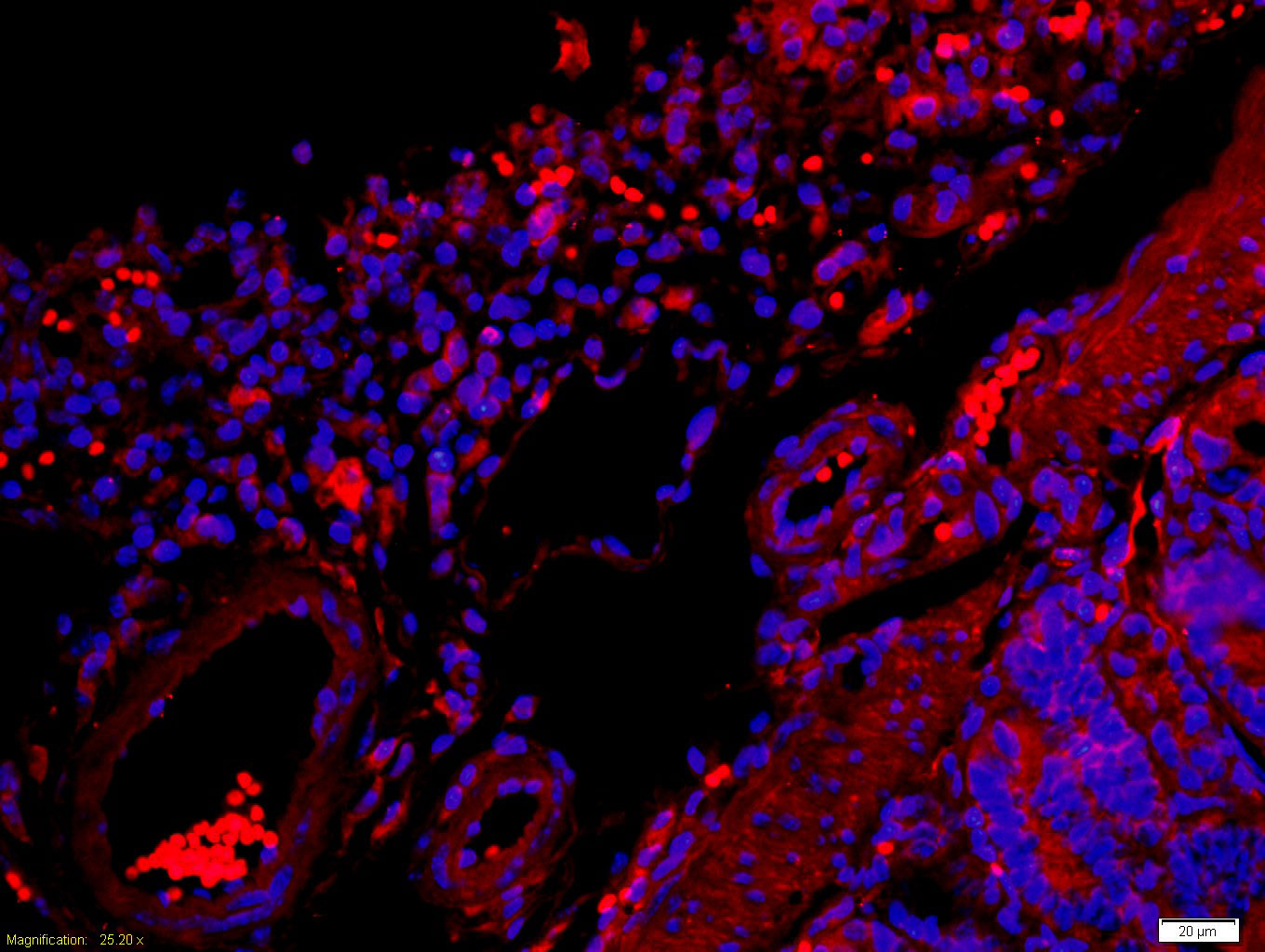 Tissue/cell: mouse intestine tissue;4% Paraformaldehyde-fixed and paraffin-embedded;
Antigen retrieval: citrate buffer ( 0.01M, pH 6.0 ), Boiling bathing for 15min; Blocking buffer (normal goat serum,C-0005) at 37℃ for 20 min;
Incubation: Anti-Caspase-6 Polyclonal Antibody, Unconjugated(SL0084R) 1:200, overnight at 4°C; The secondary antibody was Goat Anti-Rabbit IgG, Cy3 conjugated(SL0295G-Cy3)used at 1:200 dilution for 40 minutes at 37°C. DAPI(5ug/ml,blue,C-0033) was used to stain the cell nuclei
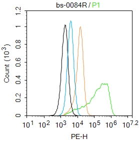 Blank control:U-2OS.
Primary Antibody (green line): Rabbit Anti-Caspase-6 subunit p18 antibody (SL0084R)
Dilution: 2μg /10^6 cells;
Isotype Control Antibody (orange line): Rabbit IgG .
Secondary Antibody : Goat anti-rabbit IgG-PE
Dilution: 1μg /test.
Protocol
The cells were fixed with 4% PFA (10min at room temperature)and then permeabilized with 0.1% PBST for 20 min at room temperature. The cells were then incubated in 5%BSA to block non-specific protein-protein interactions for 30 min at room temperature .Cells stained with Primary Antibody for 30 min at room temperature. The secondary antibody used for 40 min at room temperature. Acquisition of 20,000 events was performed.
Cartpieces
Totalgoods,subtotals:¥Checkout
Partial purchase records(bought amounts latest0)
No one bought this product
User Comment(Total0User Comment Num)
- No comment


 +86 571 56623320


