
Rabbit Anti-Cytochrome C antibody
CytC; CYC; CYCS; Cytochrome c somatic; HCS; CYC_HUMAN; Cytochrome-c; MSA06; THC4.
View History [Clear]
Details
Product Name Cytochrome C Chinese Name 细胞色素C抗体 Alias CytC; CYC; CYCS; Cytochrome c somatic; HCS; CYC_HUMAN; Cytochrome-c; MSA06; THC4. literatures Research Area Tumour Cardiovascular Cell biology Neurobiology Signal transduction Apoptosis Lipoprotein The new supersedes the old Mitochondrion Immunogen Species Rabbit Clonality Polyclonal React Species Human, Mouse, Rat, (predicted: Chicken, Pig, Cow, Horse, Rabbit, Guinea Pig, ) Applications WB=1:500-2000 ELISA=1:5000-10000 IHC-P=1:100-500 IHC-F=1:100-500 Flow-Cyt=1μg/Test ICC=1:100-500 IF=1:100-500 (Paraffin sections need antigen repair)
not yet tested in other applications.
optimal dilutions/concentrations should be determined by the end user.Theoretical molecular weight 12kDa Detection molecular weight 14.4kDa Cellular localization cytoplasmic The cell membrane Mitochondrion Form Liquid Concentration 1mg/ml immunogen KLH conjugated synthetic peptide derived from human Cytochrome C: 51-105/105 Lsotype IgG Purification affinity purified by Protein A Buffer Solution 0.01M TBS(pH7.4) with 1% BSA, 0.03% Proclin300 and 50% Glycerol. Storage Shipped at 4℃. Store at -20 °C for one year. Avoid repeated freeze/thaw cycles. Attention This product as supplied is intended for research use only, not for use in human, therapeutic or diagnostic applications. PubMed PubMed Product Detail Cytochrome C is an electron transporting protein that resides within the intermembrane space of the mitochondria, where it plays a critical role in the process of oxidative phosphorylation and production of cellular ATP. An increasing amount of interest has been directed toward the role which cytocrome C has been demonstrated to play in apoptotic processes. Following exposure to apoptotic stimuli, cytochrome C is rapidly released from the mitochondria into the cytosol, an event which may be required for the completion of apoptosis in some systems. Cytosolic cytochrome C functions in the activation of caspase 3, an ICE family molecule that is a key effector of apoptosis.
Function:
Electron carrier protein. The oxidized form of the cytochrome c heme group can accept an electron from the heme group of the cytochrome c1 subunit of cytochrome reductase. Cytochrome c then transfers this electron to the cytochrome oxidase complex, the final protein carrier in the mitochondrial electron-transport chain.
Plays a role in apoptosis. Suppression of the anti-apoptotic members or activation of the pro-apoptotic members of the Bcl-2 family leads to altered mitochondrial membrane permeability resulting in release of cytochrome c into the cytosol. Binding of cytochrome c to Apaf-1 triggers the activation of caspase-9, which then accelerates apoptosis by activating other caspases.
Subcellular Location:
Mitochondrion intermembrane space. Note=Loosely associated with the inner membrane.
Post-translational modifications:
Binds 1 heme group per subunit.
Phosphorylation at Tyr-49 and Tyr-98 both reduce by half the turnover in the reaction with cytochrome c oxidase, down-regulating mitochondrial respiration.
DISEASE:
Defects in CYCS are the cause of thrombocytopenia type 4 (THC4) [MIM:612004]; also known as autosomal dominant thrombocytopenia type 4. Thrombocytopenia is the presence of relatively few platelets in blood. THC4 is a non-syndromic form of thrombocytopenia. Clinical manifestations of thrombocytopenia are absent or mild. THC4 may be caused by dysregulated platelet formation.
Similarity:
Belongs to the cytochrome c family.
SWISS:
P99999
Gene ID:
54205
Database links:Entrez Gene: 54205 Human
Entrez Gene: 13063 Mouse
Omim: 123970 Human
SwissProt: P99999 Human
SwissProt: P62897 Mouse
Unigene: 437060 Human
细胞色素C(cytC)是一种电子传递链蛋白为Mitochondrion呼吸链必须的成份之一。在哺乳动物细胞中,如此高度保守性蛋白常分布在Mitochondrion内膜。
新近研究证明cytoplasmic中细胞色素C为激活细胞调亡所必需的因子。在调亡的过程中,细胞色素C从Mitochondrion膜被易位到cytoplasmic,由细胞色素C激活Caspase-3(CPP32)。
细胞色素C的易位可被过量表达的Bcl-2阻断。细胞色素B与细胞色素C1和Rieske蛋白相结合而形成复合物III(也称细胞色素B-C1复合物)参与细胞呼吸链。该蛋白动物种属间同源性较高;如 :猪、犬、牛、鸡、豚鼠等。
Product Picture
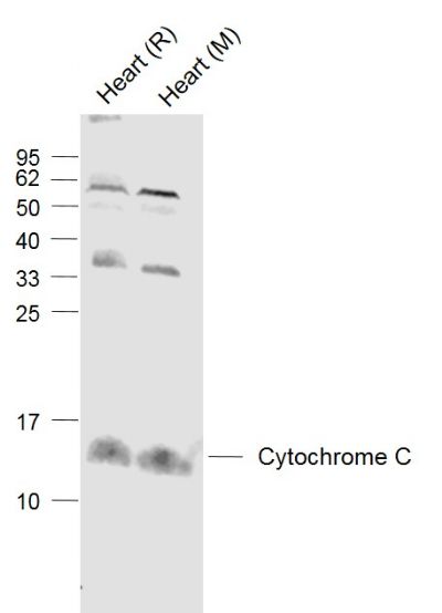
Lane 1: Heart (Rat) Lysate at 40 ug
Lane 2: Heart (Mouse) Lysate at 40 ug
Primary: Anti-Cytochrome C (SL0013R) at 1/1000 dilution
Secondary: IRDye800CW Goat Anti-Rabbit IgG at 1/20000 dilution
Predicted band size: 14.4 kD
Observed band size: 14.4 kD
Sample:
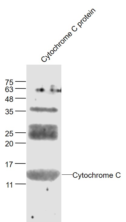
Cytochrome C protein at 30 ug
Primary: Anti- Cytochrome C (SL0013R) at 1/300 dilution
Secondary: IRDye800CW Goat Anti-Rabbit IgG at 1/20000 dilution
Predicted band size: 14 kD
Observed band size: 14 kD
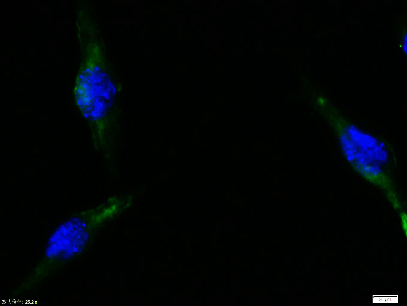
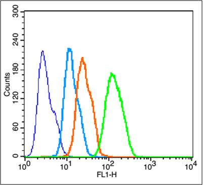
Tissue/cell:Sh-sy5y cell; 4% Paraformaldehyde-fixed; Triton X-100 at room temperature for 20 min; Blocking buffer (normal goat serum, C-0005) at 37°C for 20 min; Antibody incubation with (Cytochrome C) polyclonal Antibody, Unconjugated (SL0013R) 1:100, 90 minutes at 37°C; followed by a FITC conjugated Goat Anti-Rabbit IgG antibody at 37°C for 90 minutes, DAPI (blue, C02-04002) was used to stain the cell nuclei.Tissue/cell:Sh-sy5y cell; 4% Paraformaldehyde-fixed; Triton X-100 at room temperature for 20 min; Blocking buffer (normal goat serum, C-0005) at 37°C for 20 min; Antibody incubation with (Cytochrome C) polyclonal Antibody, Unconjugated (SL0013R) 1:100, 90 minutes at 37°C; followed by a FITC conjugated Goat Anti-Rabbit IgG antibody at 37°C for 90 minutes, DAPI (blue, C02-04002) was used to stain the cell nuclei.Tissue/cell:Sh-sy5y cell; 4% Paraformaldehyde-fixed; Triton X-100 at room temperature for 20 min; Blocking buffer (normal goat serum, C-0005) at 37°C for 20 min; Antibody incubation with (Cytochrome C) polyclonal Antibody, Unconjugated (SL0013R) 1:100, 90 minutes at 37°C; followed by a FITC conjugated Goat Anti-Rabbit IgG antibody at 37°C for 90 minutes, DAPI (blue, C02-04002) was used to stain the cell nuclei.Blank control: HepG2(blue).
Primary Antibody:Rabbit Anti-Cytochrome C antibody (SL0013R,Green); Dilution: 1μg in 100 μL 1X PBS containing 0.5% BSA;
Isotype Control Antibody: Rabbit IgG(orange) ,used under the same conditions;
Secondary Antibody: Goat anti-rabbit IgG-FITC(white blue), Dilution: 1:200 in 1 X PBS containing 0.5% BSA.
Protocol
The cells were fixed with 2% paraformaldehyde for 10 min at 37℃. Primary antibody (SL0013R, 1μg /1x10^6 cells) were incubated for 30 min at room temperature, followed by 1 X PBS containing 0.5% BSA + 1 0% goat serum (15 min) to block non-specific protein-protein interactions. Then the Goat Anti-rabbit IgG/FITC antibody was added into the blocking buffer mentioned above to react with the primary antibody at 1/200 dilution for 40 min at room temperature. Acquisition of 20,000 events was performed.
Partial purchase records(bought amounts latest0)
No one bought this product
User Comment(Total0User Comment Num)
- No comment


 +86 571 56623320


