
Rabbit Anti-Peroxiredoxin 1 antibody
Peroxiredoxin-1; Peroxiredoxin 1; Prdx 1; Prdx1; PRDX1_HUMAN; Hbp23; Heme binding 23 kDa protein; Macrophage 23 Kd stress protein; Macrophage 23kDa stress protein; Macrophase stress protein 22kDa; Macrophase stress protein 23 kd; MSP23; Natural killer cel
View History [Clear]
Details
Product Name Peroxiredoxin 1 Chinese Name 过氧化还原酶1抗体 Alias Peroxiredoxin-1; Peroxiredoxin 1; Prdx 1; Prdx1; PRDX1_HUMAN; Hbp23; Heme binding 23 kDa protein; Macrophage 23 Kd stress protein; Macrophage 23kDa stress protein; Macrophase stress protein 22kDa; Macrophase stress protein 23 kd; MSP23; Natural killer cell enhancing factor A; Natural killer cell-enhancing factor A; Natural killer enhancing factor A; NKEF A; NKEF-A; NkefA; OSF3; Osteoblast specific factor 3; PAG; Paga; PAGB; Proliferation associated gene A; Proliferation associated protein PAG; Proliferation-associated gene protein; PrxI; Tdpx 2; Tdpx2; TDX2; Thioredoxin dependent peroxide reductase 2; Thioredoxin peroxidase 2; Thioredoxin-dependent peroxide reductase 2; TPxA; Trx dependent peroxide reductase 2. literatures Research Area Tumour Cell biology immunology Immunogen Species Rabbit Clonality Polyclonal React Species Human, Mouse, Rat, (predicted: Dog, Cow, Horse, Rabbit, ) Applications WB=1:500-2000 ELISA=1:5000-10000 IHC-P=1:100-500 IHC-F=1:100-500 Flow-Cyt=2μg/Test IF=1:100-500 (Paraffin sections need antigen repair)
not yet tested in other applications.
optimal dilutions/concentrations should be determined by the end user.Theoretical molecular weight 22kDa Cellular localization cytoplasmic The cell membrane Form Liquid Concentration 1mg/ml immunogen KLH conjugated synthetic peptide derived from human PRDX1: 1-100/199 Lsotype IgG Purification affinity purified by Protein A Buffer Solution 0.01M TBS(pH7.4) with 1% BSA, 0.03% Proclin300 and 50% Glycerol. Storage Shipped at 4℃. Store at -20 °C for one year. Avoid repeated freeze/thaw cycles. Attention This product as supplied is intended for research use only, not for use in human, therapeutic or diagnostic applications. PubMed PubMed Product Detail This gene encodes a member of the peroxiredoxin family of antioxidant enzymes, which reduce hydrogen peroxide and alkyl hydroperoxides. The encoded protein may play an antioxidant protective role in cells, and may contribute to the antiviral activity of CD8(+) T-cells. This protein may have a proliferative effect and play a role in cancer development or progression. Four transcript variants encoding the same protein have been identified for this gene. [provided by RefSeq, Jan 2011].
Function:
Involved in redox regulation of the cell. Reduces peroxides with reducing equivalents provided through the thioredoxin system but not from glutaredoxin. May play an important role in eliminating peroxides generated during metabolism. Might participate in the signaling cascades of growth factors and tumor necrosis factor-alpha by regulating the intracellular concentrations of H(2)O(2). Reduces an intramolecular disulfide bond in GDPD5 that gates the ability to GDPD5 to drive postmitotic motor neuron differentiation.
Subunit:
Homodimer; disulfide-linked, upon oxidation. May form heterodimers with AOP2. Interacts with GDPD5; forms a mixed-disulfide with GDPD5 (By similarity).
Subcellular Location:
Cytoplasm. Melanosome.
Post-translational modifications:
Phosphorylated on Thr-90 during the M-phase, which leads to a more than 80% decrease in enzymatic activity.
Similarity:
Belongs to the AhpC/TSA family.
Contains 1 thioredoxin domain.
SWISS:
Q06830
Gene ID:
5052
Database links:
Entrez Gene: 5052 Human
Entrez Gene: 18477 Mouse
Omim: 176763 Human
SwissProt: Q9JKY1 Chinese Hamster
SwissProt: Q06830 Human
SwissProt: P35700 Mouse
Unigene: 180909 Human
Unigene: 30929 Mouse
Unigene: 2845 Rat
该蛋白定位于细胞质。Product Picture 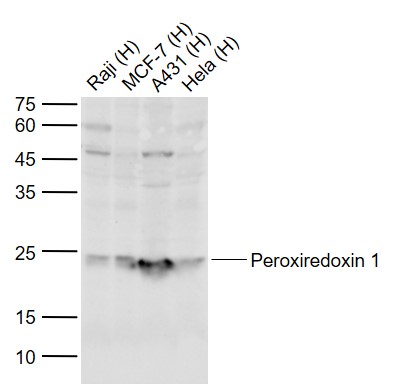 Sample:
Lane 1: Human Raji cell lysates
Lane 2: Human Hela cell lysates
Lane 3: Human A431 cell lysates
Lane 4: Human Hela cell lysates
Primary: Anti-Peroxiredoxin 1 (SL3875R) at 1/1000 dilution
Secondary: IRDye800CW Goat Anti-Rabbit IgG at 1/20000 dilution
Predicted band size: 22 kD
Observed band size: 23 kD
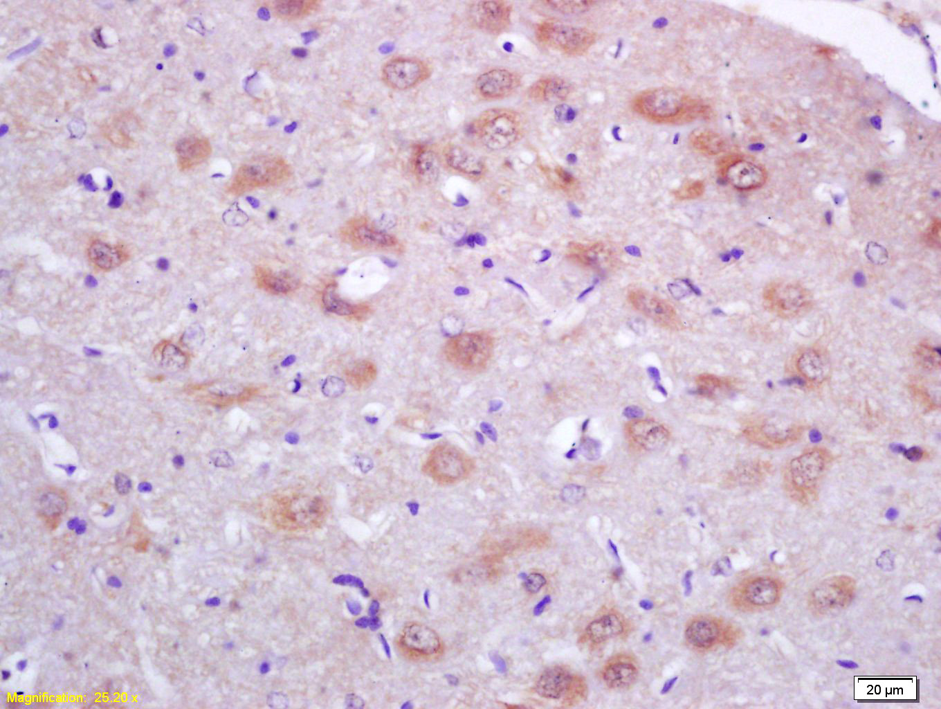 Tissue/cell: Rat brain; 4% Paraformaldehyde-fixed and paraffin-embedded;
Antigen retrieval: citrate buffer ( 0.01M, pH 6.0 ), Boiling bathing for 15min; Block endogenous peroxidase by 3% Hydrogen peroxide for 30min; Blocking buffer (normal goat serum,C-0005) at 37℃ for 20 min;
Incubation: Anti-PRDX1 Polyclonal Antibody, Unconjugated(SL3875R) 1:200, overnight at 4°C, followed by conjugation to the secondary antibody(SP-0023) and DAB(C-0010) staining
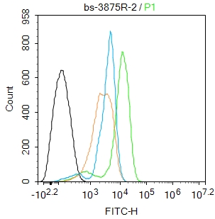 Blank control: Hela.
Primary Antibody (green line): Rabbit Anti-Peroxiredoxin 1 antibody (SL3875R)
Dilution: 2ug/Test;
Secondary Antibody : Goat anti-rabbit IgG-FITC
Dilution: 0.5ug/Test.
Protocol
The cells were fixed with 4% PFA (10min at room temperature)and then permeabilized with 0.1%PBST for 20 min at room temperature.The cells were then incubated in 5%BSA to block non-specific protein-protein interactions for 30 min at room temperature .Cells stained with Primary Antibody for 30 min at room temperature. The secondary antibody used for 40 min at room temperature. Acquisition of 20,000 events was performed.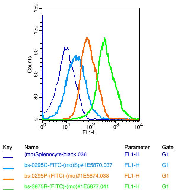 Blank control: mouse splenocytes(blue)
Isotype Control Antibody: Rabbit IgG(orange) ; Secondary Antibody: Goat anti-rabbit IgG-FITC(white blue), Dilution: 1:100 in 1 X PBS containing 0.5% BSA ; Primary Antibody Dilution: 1μl in 100 μL1X PBS containing 0.5% BSA(green).
Cartpieces
Totalgoods,subtotals:¥Checkout
Bought notes(bought amounts latest0)
No one bought this product
User Comment(Total0User Comment Num)
- No comment


 +86 571 56623320


