
Rabbit Anti-Claudin 5 antibody
AWAL; BEC 1; BEC1; Claudin 5 (transmembrane protein deleted in velocardiofacial syndrome); Claudin5; Claudin-5;CLDN 5; CLDN5; CPETR L1; CPETRL 1; CPETRL1; TMDVCF; TMVCF; Transmembrane protein deleted in VCFS; Transmembrane protein deleted in velocardiofac
View History [Clear]
Details
Product Name Claudin 5 Chinese Name 紧密连接蛋白5抗体 Alias AWAL; BEC 1; BEC1; Claudin 5 (transmembrane protein deleted in velocardiofacial syndrome); Claudin5; Claudin-5;CLDN 5; CLDN5; CPETR L1; CPETRL 1; CPETRL1; TMDVCF; TMVCF; Transmembrane protein deleted in VCFS; Transmembrane protein deleted in velocardiofacial syndrome; Androgen withdrawal and apoptosis induced protein RVP1 like; CLD5_HUMAN. literatures Research Area Signal transduction Cell adhesion molecule Cytoskeleton Immunogen Species Rabbit Clonality Polyclonal React Species Human, Mouse, (predicted: Rat, Pig, Cow, Horse, Rabbit, Sheep, ) Applications WB=1:500-2000 ELISA=1:5000-10000 IHC-P=1:100-500 Flow-Cyt=2ug/Test (Paraffin sections need antigen repair)
not yet tested in other applications.
optimal dilutions/concentrations should be determined by the end user.Theoretical molecular weight 23kDa Cellular localization The cell membrane Extracellular matrix Form Liquid Concentration 1mg/ml immunogen KLH conjugated synthetic peptide derived from human Claudin 5: 29-81/218 <Extracellular> Lsotype IgG Purification affinity purified by Protein A Buffer Solution 0.01M TBS(pH7.4) with 1% BSA, 0.03% Proclin300 and 50% Glycerol. Storage Shipped at 4℃. Store at -20 °C for one year. Avoid repeated freeze/thaw cycles. Attention This product as supplied is intended for research use only, not for use in human, therapeutic or diagnostic applications. PubMed PubMed Product Detail This gene encodes a member of the claudin family. Claudins are integral membrane proteins and components of tight junction strands. Tight junction strands serve as a physical barrier to prevent solutes and water from passing freely through the paracellular space between epithelial or endothelial cell sheets. Mutations in this gene have been found in patients with velocardiofacial syndrome. Alternatively spliced transcript variants encoding the same protein have been found for this gene. [provided by RefSeq, Aug 2008]
Function:
Plays a major role in tight junction-specific obliteration of the intercellular space.
Subunit:
Directly interacts with TJP1/ZO-1, TJP2/ZO-2 and TJP3/ZO-3. Interacts with MPDZ.
Subcellular Location:
Cell junction, tight junction. Cell membrane; Multi-pass membrane protein.
Tissue Specificity:
Expressed in activated, but not resting, CD4+ T-cells and activated monocytes.
Similarity:
Belongs to the claudin family.
SWISS:
O00501
Gene ID:
7122
Database links:Entrez Gene: 7122 Human
Entrez Gene: 12741 Mouse
Omim: 602101 Human
SwissProt: O00501 Human
SwissProt: O54942 Mouse
Unigene: 505337 Human
Unigene: 22768 Mouse
Product Picture 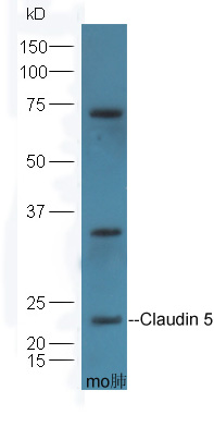 Protein: lung(mouse) lysates at 45ug; Primary: Anti-Claudin 5 (SL10296R) at 1:300;
Secondary: HRP conjugated Goat-Anti-Rabbit IgG(bse-0295G-HRP) at 1: 5000;
ECL excitated the fluorescence;
Predicted band size :23 kD
Observed band size :23kD
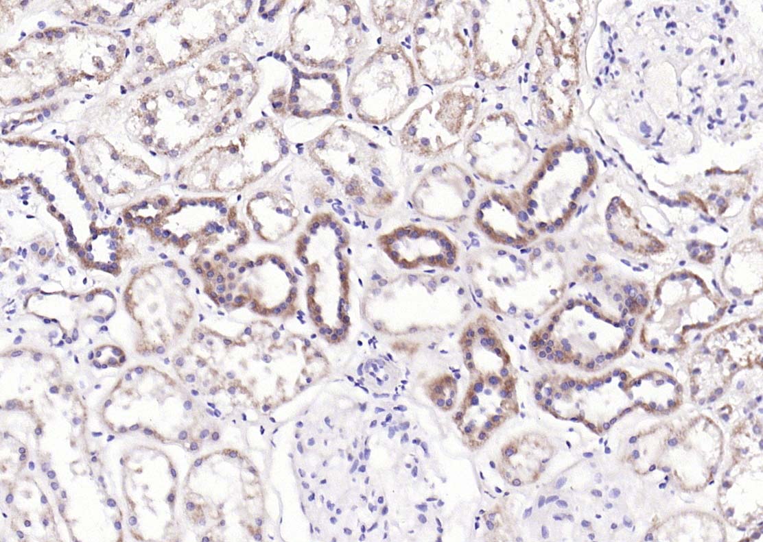 Paraformaldehyde-fixed, paraffin embedded (Human kidney); Antigen retrieval by boiling in sodium citrate buffer (pH6.0) for 15min; Block endogenous peroxidase by 3% hydrogen peroxide for 20 minutes; Blocking buffer (normal goat serum) at 37°C for 30min; Antibody incubation with (Claudin 5) Polyclonal Antibody, Unconjugated (SL10296R) at 1:200 overnight at 4°C, followed by operating according to SP Kit(Rabbit) (sp-0023) instructionsand DAB staining.
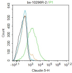 Blank control: K562.
Primary Antibody (green line): Rabbit Anti-Claudin 5 antibody (SL10296R)
Dilution:2μg /10^6 cells;
Isotype Control Antibody (orange line): Rabbit IgG .
Secondary Antibody : Goat anti-rabbit IgG-FITC
Dilution: 0.5μg /test.
Protocol
The cells were incubated in 5%BSA to block non-specific protein-protein interactions for 30 min at room temperature .Cells stained with Primary Antibody for 30 min at room temperature. The secondary antibody used for 40 min at room temperature. Acquisition of 20,000 events was performed.
Cartpieces
Totalgoods,subtotals:¥Checkout
Bought notes(bought amounts latest0)
No one bought this product
User Comment(Total0User Comment Num)
- No comment


 +86 571 56623320
 +86 18668110335

