
Rabbit Anti-EphB2 antibody
PHB2_HUMAN; Ephrin type-B receptor 2; EC:2.7.10.1; Developmentally-regulated Eph-related tyrosine kinase; ELK-related tyrosine kinase; EPH tyrosine kinase 3; EPH-like kinase 5 (EK5; hEK5); Renal carcinoma antigen NY-REN-47; Tyrosine-protein kinase TYRO5;
View History [Clear]
Details
Product Name EphB2 Chinese Name 酪氨酸蛋白激酶受体B2抗体 Alias PHB2_HUMAN; Ephrin type-B receptor 2; EC:2.7.10.1; Developmentally-regulated Eph-related tyrosine kinase; ELK-related tyrosine kinase; EPH tyrosine kinase 3; EPH-like kinase 5 (EK5; hEK5); Renal carcinoma antigen NY-REN-47; Tyrosine-protein kinase TYRO5; Tyrosine-protein kinase receptor EPH-3; EphB2/CTF1; EphB2/CTF2; DRT; EPHT3; EPTH3; TYRO5; CAPB; PCBC; BDPLT22; literatures Research Area Tumour Cell biology immunology Signal transduction transcriptional regulatory factor Kinases and Phosphatases The cell membrane受体 Immunogen Species Rabbit Clonality Polyclonal React Species Human, Mouse, Rat, (predicted: Chicken, Dog, Cow, ) Applications WB=1:500-2000 ELISA=1:5000-10000 IHC-P=1:100-500 IHC-F=1:100-500 Flow-Cyt=3ug/Test IF=1:100-500 (Paraffin sections need antigen repair)
not yet tested in other applications.
optimal dilutions/concentrations should be determined by the end user.Theoretical molecular weight 108kDa Cellular localization cytoplasmic The cell membrane Form Liquid Concentration 1mg/ml immunogen KLH conjugated synthetic peptide derived from human EPHB2: 551-650/1055 Lsotype IgG Purification affinity purified by Protein A Buffer Solution 0.01M TBS(pH7.4) with 1% BSA, 0.03% Proclin300 and 50% Glycerol. Storage Shipped at 4℃. Store at -20 °C for one year. Avoid repeated freeze/thaw cycles. Attention This product as supplied is intended for research use only, not for use in human, therapeutic or diagnostic applications. PubMed PubMed Product Detail This gene encodes a member of the Eph receptor family of receptor tyrosine kinase transmembrane glycoproteins. These receptors are composed of an N-terminal glycosylated ligand-binding domain, a transmembrane region and an intracellular kinase domain. They bind ligands called ephrins and are involved in diverse cellular processes including motility, division, and differentiation. A distinguishing characteristic of Eph-ephrin signaling is that both receptors and ligands are competent to transduce a signaling cascade, resulting in bidirectional signaling. This protein belongs to a subgroup of the Eph receptors called EphB. Proteins of this subgroup are distinguished from other members of the family by sequence homology and preferential binding affinity for membrane-bound ephrin-B ligands. Allelic variants are associated with prostate and brain cancer susceptibility. Alternative splicing results in multiple transcript variants. [provided by RefSeq, May 2015]
Function:
Receptor for members of the ephrin-B family. Phosphorylates ARHGEF15, leading to its ubiquitination and degradation by the proteasome which promotes EFNB1-dependent synapse formation. Can function in aspects of retinal ganglion cell axon guidance to the optic disk even when lacking its tyrosine kinase domain. Acts as a tumor suppressor.
Subunit:
Heterotetramer upon binding of the ligand. The heterotetramer is composed of an ephrin dimer and a receptor dimer. Oligomerization is probably required to induce biological responses. Interacts (via PDZ-binding motif) with GRIP1 and PICK1 (via PDZ domain). Interacts with ARHGEF15; mediates ARHGEF15 phosphorylation, ubiquitination and degradation by the proteasome. Interacts with AQP1; involved in endolymph production in the inner ear.
Subcellular Location:
Cell membrane; Single-pass type I membrane protein. Cell projection, axon. Cell projection, dendrite.
Tissue Specificity:
Brain, heart, lung, kidney, placenta, pancreas, liver and skeletal muscle. Preferentially expressed in fetal brain.
DISEASE:
Defects in EPHB2 may be a cause of susceptibility to prostate cancer (PC) [MIM:176807]. It is a malignancy originating in tissues of the prostate. Most prostate cancers are adenocarcinomas that develop in the acini of the prostatic ducts. Other rare histopathologic types of prostate cancer that occur in approximately 5% of patients include small cell carcinoma, mucinous carcinoma, prostatic ductal carcinoma, transitional cell carcinoma, squamous cell carcinoma, basal cell carcinoma, adenoid cystic carcinoma (basaloid), signet-ring cell carcinoma and neuroendocrine carcinoma. Note=EPHB2 mutations have been found in a prostate cancer cell line derived from a brain metastasis.
Similarity:
Belongs to the protein kinase superfamily. Tyr protein kinase family. Ephrin receptor subfamily.
Contains 2 fibronectin type-III domains.
Contains 1 protein kinase domain.
Contains 1 SAM (sterile alpha motif) domain.
SWISS:
P29323
Gene ID:
2048
Database links:Entrez Gene: 2048 Human
Entrez Gene: 13844 Mouse
Omim: 600997 Human
SwissProt: P29323 Human
SwissProt: P54763 Mouse
Unigene: 523329 Human
Unigene: 250981 Mouse
Product Picture 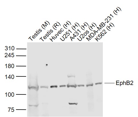 Sample:
Lane 1: Testis (Mouse) Lysate at 40 ug
Lane 2: Testis (Rat) Lysate at 40 ug
Lane 3: Huvec (Human) Cell Lysate at 30 ug
Lane 4: U251 (Human) Cell Lysate at 30 ug
Lane 5: A431 (Human) Cell Lysate at 30 ug
Lane 6: U2os (Human) Cell Lysate at 30 ug
Lane 7: MDA-MB-231 (Human) Cell Lysate at 30 ug
Lane 8: K562 (Human) Cell Lysate at 30 ug
Primary: Anti-EphB2 (SL0996R) at 1/1000 dilution
Secondary: IRDye800CW Goat Anti-Rabbit IgG at 1/20000 dilution
Predicted band size: 125 kD
Observed band size: 120 kD
 Blank control: A431.
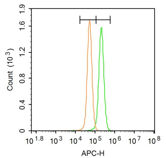
Primary Antibody (green line): Rabbit Anti-EphB2 antibody (SL0996R)
Dilution: 3μg /10^6 cells;
Isotype Control Antibody (orange line): Rabbit IgG .
Secondary Antibody: Goat anti-rabbit IgG-AF647
Dilution: 3μg /test.
Protocol
The cells were fixed with 4% PFA (10min at room temperature)and then permeabilized with 20% PBST for 20 min at room temperature. The cells were then incubated in 5%BSA to block non-specific protein-protein interactions for 30 min at room temperature .Cells stained with Primary Antibody for 30 min at room temperature. The secondary antibody used for 40 min at room temperature. Acquisition of 20,000 events was performed.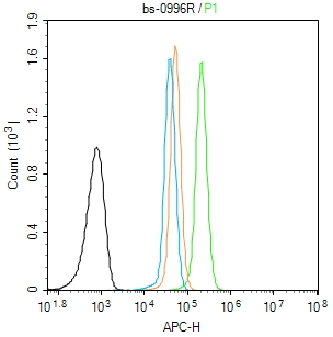 Blank control (Black line): A431 (Black).
Primary Antibody (green line): Rabbit Anti-EphB2 antibody (SL0996R)
Dilution: 1μg /10^6 cells;
Isotype Control Antibody (orange line): Rabbit IgG .
Secondary Antibody (white blue line): Goat anti-rabbit IgG-AF647
Dilution: 1μg /test.
Protocol
The cells were fixed with 4% PFA (10min at room temperature)and then permeabilized with 0.1% PBST for 20 min at room temperature. The cells were then incubated in 5%BSA to block non-specific protein-protein interactions for 30 min at room temperature.Cells stained with Primary Antibody for 30 min at room temperature. The secondary antibody used for 40 min at room temperature. Acquisition of 20,000 events was performed.Blank control (Black line): A431 (Black).
Primary Antibody (green line): Rabbit Anti-EphB2 antibody (SL0996R)
Dilution: 1μg /10^6 cells;
Isotype Control Antibody (orange line): Rabbit IgG .
Secondary Antibody (white blue line): Goat anti-rabbit IgG-AF647
Dilution: 1μg /test.
Protocol
The cells were fixed with 4% PFA (10min at room temperature)and then permeabilized with 0.1% PBST for 20 min at room temperature. The cells were then incubated in 5%BSA to block non-specific protein-protein interactions for 30 min at room temperature.Cells stained with Primary Antibody for 30 min at room temperature. The secondary antibody used for 40 min at room temperature. Acquisition of 20,000 events was performed.
Cartpieces
Totalgoods,subtotals:¥Checkout
Bought notes(bought amounts latest0)
No one bought this product
User Comment(Total0User Comment Num)
- No comment


 +86 571 56623320
 +86 18668110335

