
Rabbit Anti-CEA antibody
Carcinoembryonic antigen; Carcinoembryonic antigen related cell adhesion molecule 5; Carcinoembryonic antigen-related cell adhesion molecule 5; CD66e; CD66e antigen; CEACAM5; CEAM5_HUMAN; DKFZp781M2392; Meconium antigen 100.
View History [Clear]
Details
Product Name CEA Chinese Name 癌胚抗原抗体 Alias Carcinoembryonic antigen; Carcinoembryonic antigen related cell adhesion molecule 5; Carcinoembryonic antigen-related cell adhesion molecule 5; CD66e; CD66e antigen; CEACAM5; CEAM5_HUMAN; DKFZp781M2392; Meconium antigen 100. literatures Research Area Tumour Signal transduction Immunogen Species Rabbit Clonality Polyclonal React Species Human, Applications ELISA=1:5000-10000 IHC-P=1:100-500 IHC-F=1:100-500 Flow-Cyt=1μg/test IF=1:100-500 (Paraffin sections need antigen repair)
not yet tested in other applications.
optimal dilutions/concentrations should be determined by the end user.Theoretical molecular weight 150-200kDa Cellular localization The cell membrane Form Liquid Concentration 1mg/ml immunogen KLH conjugated synthetic peptide derived from human CEA/CD66e/CEACAM5: 301-400/702 Lsotype IgG Purification affinity purified by Protein A Buffer Solution 0.01M TBS(pH7.4) with 1% BSA, 0.03% Proclin300 and 50% Glycerol. Storage Shipped at 4℃. Store at -20 °C for one year. Avoid repeated freeze/thaw cycles. Attention This product as supplied is intended for research use only, not for use in human, therapeutic or diagnostic applications. PubMed PubMed Product Detail CEA-related cell adhesion molecules (CEACAM) belong to the carcinoembryonic antigen (CEA) family. It consists of seven CEACAM (CEACAM 1, CEACAM 3-CEACAM 8) and 11 pregnancy-specific glyco-protein (PSG 1-PSG 11) members. The CEA family proteins belong to the immunoglobulin (Ig) superfamily and are composed of one Ig variable-like (IgV) and a varying number (0-6) of Ig constant-like (IgC) domains. CEACAM molecules are membrane-bound either via a transmembrane domain or a glycosyl phosphatidyl inositol (GPI) anchor. CEACAM molecules are differentially expressed in epithelial cells or in leucocytes. Over-expression of CEA/ CEACAM 5 in tumors of epithelial origin is the basis of its wide-spread use as a tumor marker. The function of CEACAM family members varies widely: they function as cell adhesion molecules, tumor suppressors, regulators of lymphocyte and dendritic cell activation, receptors of Neisseria species and other bacteria.
Function:
Cell surface glycoprotein that plays a role in cell adhesion and in intracellular signaling. Receptor for E.coli Dr adhesins.
Subunit:
Homodimer. Binding of E.coli Dr adhesins leads to dissociation of the homodimer.
Subcellular Location:
Cell membrane; Lipid-anchor, GPI-anchor.
Tissue Specificity:
Found in adenocarcinomas of endodermally derived digestive system epithelium and fetal colon.
Post-translational modifications:
Complex immunoreactive glycoprotein with a MW of 180 kDa comprising 60% carbohydrate.
Similarity:
Belongs to the immunoglobulin superfamily. CEA family.
Contains 7 Ig-like (immunoglobulin-like) domains.
SWISS:
P06731
Gene ID:
1048
Database links:Entrez Gene: 1048 Human
Omim: 114890 Human
SwissProt: P06731 Human
Unigene: 709196 Human
CEA是一种胚胎性抗原,主要存在于胎儿消化道上皮组织,胰腺和肝癌中。CEA广泛存在各种上皮性Tumour,尤其是胃肠道恶性Tumour,CEA分布阳性类型与Tumour的恶性度有关。CEA与EMA一样可作为上皮性恶性Tumour的重要标记。Product Picture 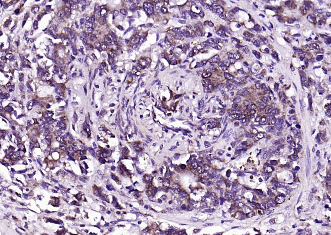 Paraformaldehyde-fixed, paraffin embedded (human rectal carcinoma); Antigen retrieval by boiling in sodium citrate buffer (pH6.0) for 15min; Block endogenous peroxidase by 3% hydrogen peroxide for 20 minutes; Blocking buffer (normal goat serum) at 37°C for 30min; Antibody incubation with (CEA) Polyclonal Antibody, Unconjugated (SL0719R) at 1:200 overnight at 4°C, followed by operating according to SP Kit(Rabbit) (sp-0023) instructionsand DAB staining.
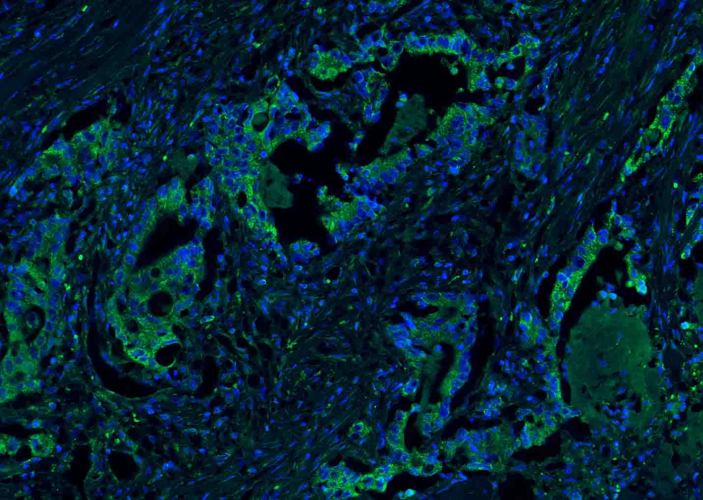 Paraformaldehyde-fixed, paraffin embedded (human rectal carcinoma); Antigen retrieval by boiling in sodium citrate buffer (pH6.0) for 15min; Blocking buffer (normal goat serum) at 37°C for 30min; Antibody incubation with (CEA) Polyclonal Antibody, Unconjugated (SL0719R) at 1:200 overnight at 4°C, followed by a conjugated Goat Anti-Rabbit IgG antibody (SL0295G-FITC) for 90 minutes, and DAPI for nuclei staining.
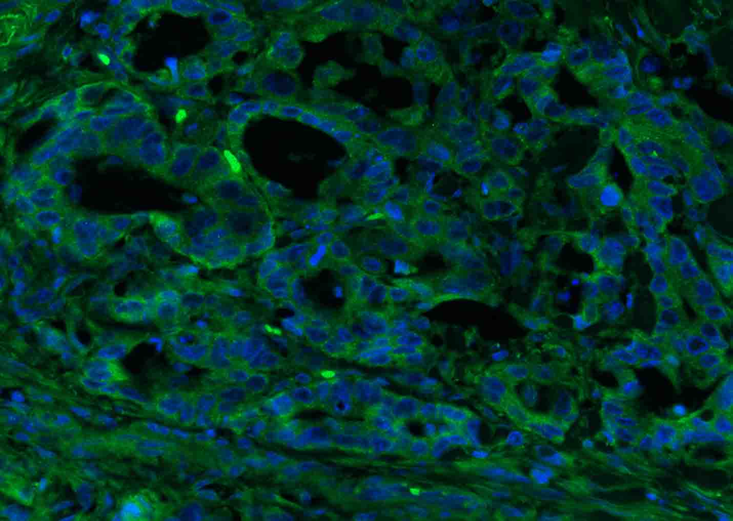 Paraformaldehyde-fixed, paraffin embedded (human colon carcinoma); Antigen retrieval by boiling in sodium citrate buffer (pH6.0) for 15min; Blocking buffer (normal goat serum) at 37°C for 30min; Antibody incubation with (CEA) Polyclonal Antibody, Unconjugated (SL0719R) at 1:200 overnight at 4°C, followed by a conjugated Goat Anti-Rabbit IgG antibody (SL0295G-FITC) for 90 minutes, and DAPI for nuclei staining.
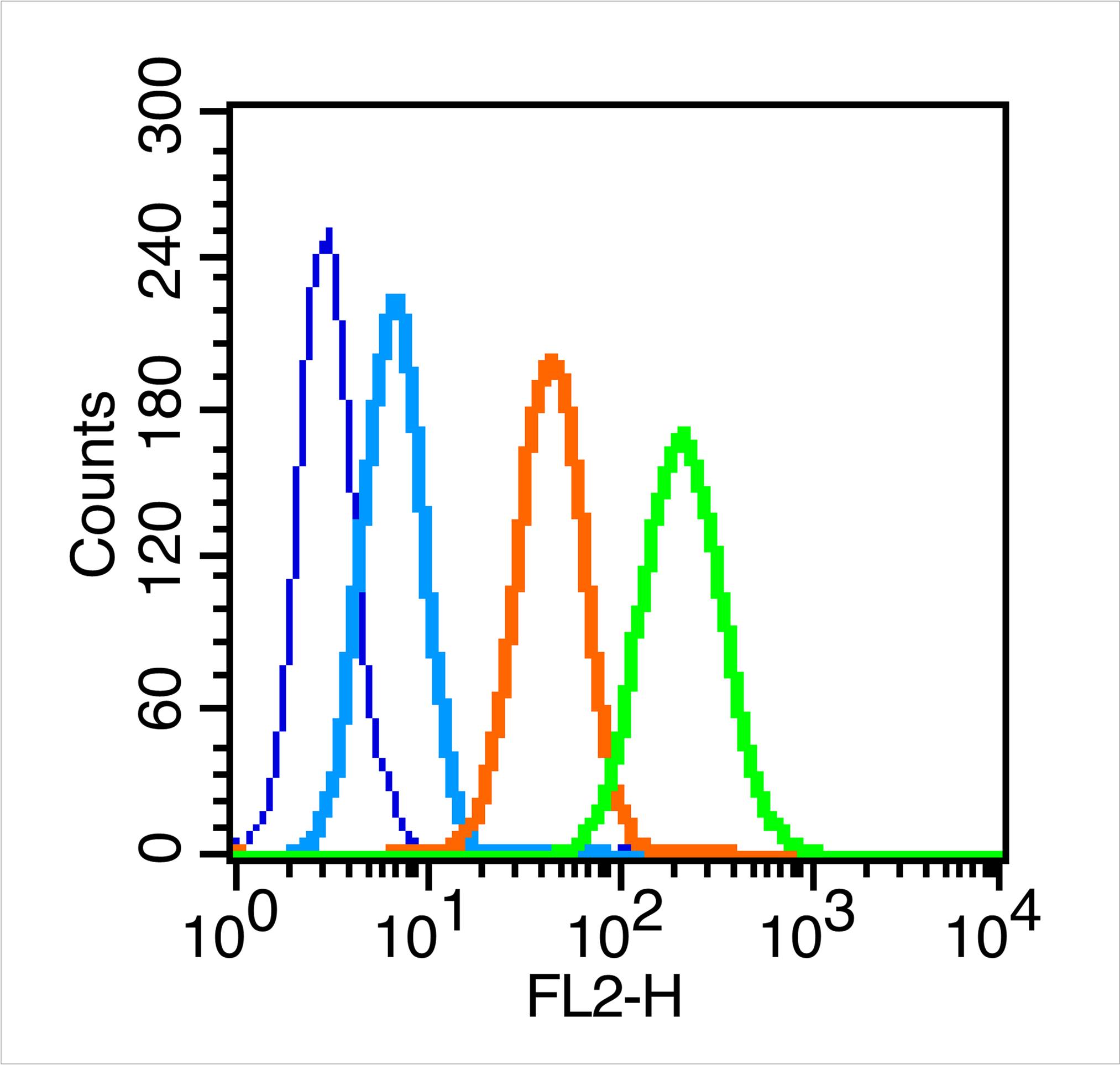 Blank control (blue line): MCF7 (fixed with 70% methanol overnight at 4℃).
Primary Antibody (green line): Rabbit Anti-CEA antibody (SL0719R)
Dilution: 0.2μg /10^6 cells;
Isotype Control Antibody (orange line): Rabbit IgG .
Secondary Antibody (white blue line): Goat anti-rabbit IgG-PE
Dilution: 1μg /test.
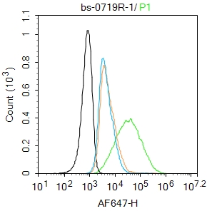 Blank control: MCF7.
Primary Antibody (green line): Rabbit Anti-CEA antibody (SL0719R)
Dilution: 1μg /10^6 cells;
Isotype Control Antibody (orange line): Rabbit IgG .
Secondary Antibody : Goat anti-rabbit IgG-AF647
Dilution: 1μg /test.
Protocol
The cells were incubated in 5%BSA to block non-specific protein-protein interactions for 30 min at room temperature .Cells stained with Primary Antibody for 30 min at room temperature. The secondary antibody used for 40 min at room temperature. Acquisition of 20,000 events was performed.
Cartpieces
Totalgoods,subtotals:¥Checkout
Bought notes(bought amounts latest0)
No one bought this product
User Comment(Total0User Comment Num)
- No comment


 +86 571 56623320
 +86 18668110335

