
Rabbit Anti-CD62p antibody
P-selectin; CD_antigen: CD62P; CD62 antigen-like family member P; GMP 140; GMP-140; GMRP; Granule membrane protein 140; Granulocyte membrane protein; GRMP; LECAM 3; LECAM3; Leukocyte endothelial cell adhesion molecule 3; Leukocyte-endothelial cell adhesio
View History [Clear]
Details
Product Name CD62p Chinese Name P选择素/白细胞endothelial cells粘附分子3抗体 Alias P-selectin; CD_antigen: CD62P; CD62 antigen-like family member P; GMP 140; GMP-140; GMRP; Granule membrane protein 140; Granulocyte membrane protein; GRMP; LECAM 3; LECAM3; Leukocyte endothelial cell adhesion molecule 3; Leukocyte-endothelial cell adhesion molecule 3; P Selectin; PADGEM; Platelet activation dependent granule-external membrane protein; PSEL; sp-selectin; LYAM3_HUMAN. literatures Research Area Cardiovascular Cell biology immunology Stem cells Cell adhesion molecule epithelial cells Immunogen Species Rabbit Clonality Polyclonal React Species Human, Rat, Pig, (predicted: Mouse, Dog, Cow, Horse, ) Applications WB=1:500-2000 ELISA=1:5000-10000 IHC-P=1:100-500 IHC-F=1:100-500 Flow-Cyt=1μg/Test IF=1:100-500 (Paraffin sections need antigen repair)
not yet tested in other applications.
optimal dilutions/concentrations should be determined by the end user.Theoretical molecular weight 88kDa Cellular localization The cell membrane Form Liquid Concentration 1mg/ml immunogen KLH conjugated synthetic peptide derived from mouse P-selectin: 701-768/768 <Cytoplasmic> Lsotype IgG Purification affinity purified by Protein A Buffer Solution 0.01M TBS(pH7.4) with 1% BSA, 0.03% Proclin300 and 50% Glycerol. Storage Shipped at 4℃. Store at -20 °C for one year. Avoid repeated freeze/thaw cycles. Attention This product as supplied is intended for research use only, not for use in human, therapeutic or diagnostic applications. PubMed PubMed Product Detail This gene encodes a 140 kDa protein that is stored in the alpha-granules of platelets and Weibel-Palade bodies of endothelial cells. This protein redistributes to the plasma membrane during platelet activation and degranulation and mediates the interaction of activated endothelial cells or platelets with leukocytes. The membrane protein is a calcium-dependent receptor that binds to sialylated forms of Lewis blood group carbohydrate antigens on neutrophils and monocytes. Alternative splice variants may occur but are not well documented. [provided by RefSeq, Jul 2008]
Function:
Ca(2+)-dependent receptor for myeloid cells that binds to carbohydrates on neutrophils and monocytes. Mediates the interaction of activated endothelial cells or platelets with leukocytes. The ligand recognized is sialyl-Lewis X. Mediates rapid rolling of leukocyte rolling over vascular surfaces during the initial steps in inflammation through interaction with PSGL1.
Subunit:
Interacts with SNX17. Interacts with PSGL1/SEPL and PODXL2 and mediates neutrophil adhesion and leukocyte rolling. This interaction requires the sialyl-Lewis X epitope of PSGL1 and PODXL2, and specific tyrosine sulfation on PSGL1.
Subcellular Location:
Membrane; Single-pass type I membrane protein.
Tissue Specificity:
Stored in the alpha-granules of platelets and Weibel-Palade bodies of endothelial cells. Upon cell activation by agonists, P-selectin is transported rapidly to the cell surface.
DISEASE:
Defects in SELP may be a cause of susceptibility to ischemic stroke (ISCHSTR) [MIM:601367]; also known as cerebrovascular accident or cerebral infarction. A stroke is an acute neurologic event leading to death of neural tissue of the brain and resulting in loss of motor, sensory and/or cognitive function. Ischemic strokes, resulting from vascular occlusion, is considered to be a highly complex disease consisting of a group of heterogeneous disorders with multiple genetic and environmental risk factors.
Similarity:
Belongs to the selectin/LECAM family.
Contains 1 C-type lectin domain.
Contains 1 EGF-like domain.
Contains 9 Sushi (CCP/SCR) domains.
SWISS:
Q01102
Gene ID:
6403
Database links:Entrez Gene: 6403 Human
Entrez Gene: 20344 Mouse
Omim: 173610 Human
SwissProt: P16109 Human
SwissProt: Q01102 Mouse
Unigene: 73800 Human
P-选择素(P—selectin)是细胞黏附分子选择素家族的主要成员,又称CD62p,可以介导各种白细胞和Tumour细胞的粘附。
CD62p又称GMP—140或PADGEM蛋白(血小板活化依赖α颗粒膜蛋白),它在血小板与单核细胞及中性粒细胞相互作用中起关键作用。Product Picture 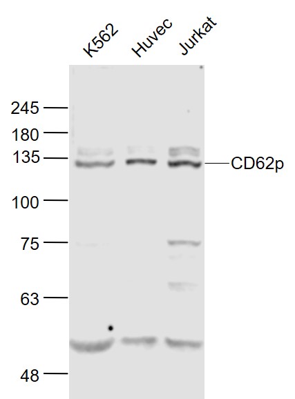 Sample:
K562(Human) Cell Lysate at 30 ug
Huvec(Human) Cell Lysate at 30 ug
Jurkat(Human) Cell Lysate at 30 ug
Primary: Anti-CD62p (SL0561R) at 1/1000 dilution
Secondary: IRDye800CW Goat Anti-Rabbit IgG at 1/20000 dilution
Predicted band size: 140'90 kD
Observed band size: 130 kD
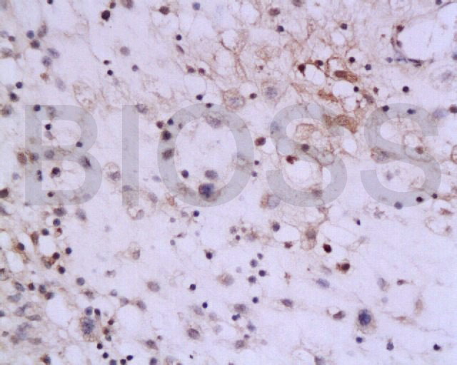 Tissue/cell: rat colon tissue; 4% Paraformaldehyde-fixed and paraffin-embedded;
Antigen retrieval: citrate buffer ( 0.01M, pH 6.0 ), Boiling bathing for 15min; Block endogenous peroxidase by 3% Hydrogen peroxide for 30min; Blocking buffer (normal goat serum,C-0005) at 37℃ for 20 min;
Incubation: Anti-P-selectin Polyclonal Antibody, Unconjugated(SL0561R) 1:200, overnight at 4°C, followed by conjugation to the secondary antibody(SP-0023) and DAB(C-0010) staining
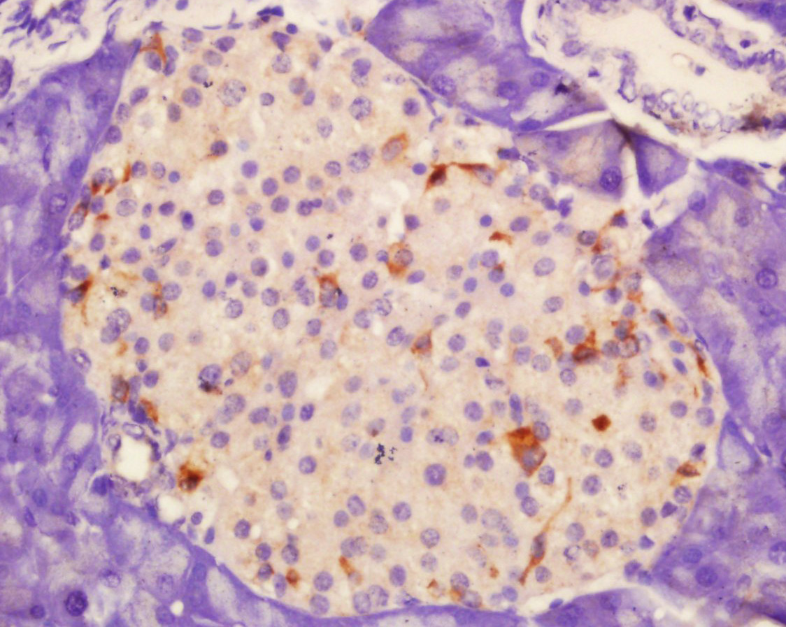 Tissue/cell: rat pancreas tissue; 4% Paraformaldehyde-fixed and paraffin-embedded;
Antigen retrieval: citrate buffer ( 0.01M, pH 6.0 ), Boiling bathing for 15min; Block endogenous peroxidase by 3% Hydrogen peroxide for 30min; Blocking buffer (normal goat serum,C-0005) at 37℃ for 20 min;
Incubation: Anti-P-selectin Polyclonal Antibody, Unconjugated(SL0561R) 1:200, overnight at 4°C, followed by conjugation to the secondary antibody(SP-0023) and DAB(C-0010) staining
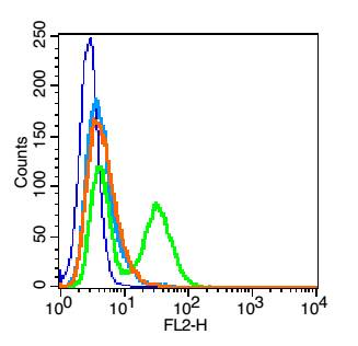 Blank control: HUVEC cells(blue).
Primary Antibody:Rabbit Anti-CD62p antibody(SL0561R), Dilution: 1μg in 100 μL 1X PBS containing 0.5% BSA;
Isotype Control Antibody: Rabbit IgG(orange) ,used under the same conditions );
Secondary Antibody: Goat anti-rabbit IgG-PE(white blue), Dilution: 1:200 in 1 X PBS containing 0.5% BSA.
Protocol
The cells were fixed with 2% paraformaldehyde (10 min) .Primary antibody (SL0561R, 1μg /1x10^6 cells) were incubated for 30 min on the ice, followed by 1 X PBS containing 0.5% BSA + 1 0% goat serum (15 min) to block non-specific protein-protein interactions. Then the Goat Anti-rabbit IgG/PE antibody was added into the blocking buffer mentioned above to react with the primary antibody at 1/200 dilution for 30 min on ice. Acquisition of 20,000 events was performed.
Cartpieces
Totalgoods,subtotals:¥Checkout
Bought notes(bought amounts latest0)
No one bought this product
User Comment(Total0User Comment Num)
- No comment


 +86 571 56623320
 +86 18668110335

